An interview with Dr Karina Serban, Assistant Professor of Medicine, National Jewish Health, conducted by April Cashin-Garbutt, MA (Cantab)
How important is exosome isolation in your research?
In the outpatient clinic and in the laboratory our current research studies aim at understanding the mechanisms of release and clearance and the biological functions of exosomes and membrane particles in the plasma of individuals who develop emphysema, a form of chronic obstructive pulmonary disease characterized by enlargement of airspaces and loss of alveoli.

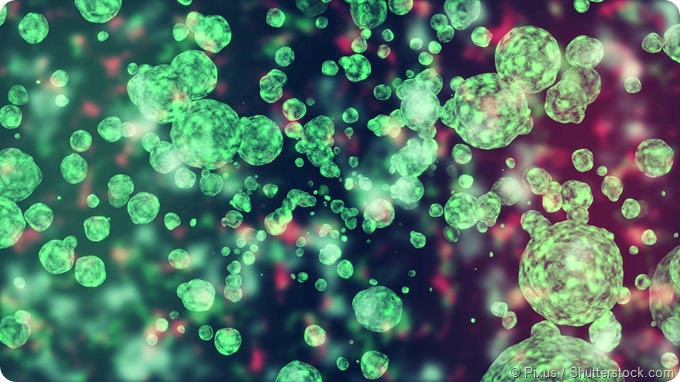
Image credit: Shutterstock
Soluble components within the cigarette smoke (CS) initially inhaled and then absorbed into the circulation are the most common injury that leads to emphysema and COPD. We have showed that acute exposure to CS is sufficient to stimulate the release of exosomes and membrane particles from lung endothelial cells with highly pro-inflammatory potential based on their cargo analysis: pro-coagulant and pro-inflammatory surface membrane molecules, bioactive lipids, and miRNAs.
The exosomes and membrane particles are extremely small, nanometer in size, circulating particles released within the bloodstream by structural, endothelial cells or circulating cells, like leukocytes or platelets. We have isolated exosomes and membrane particles using differential ultracentrifugation. This method allowed us to pellet the exosomes and membrane particles based on their density, to achieve complete separation from the fluid phase, and to resuspend the pellet in various buffer required for downstream quantitative (flow cytometry counting), qualitative (membrane proteins, lipids, miRNAs), and functional studies (uptake by macrophages).
Since not all smokers develop emphysema we believe that exosomes abundance in the blood of active smokers will allow us to identify the individuals at risk for lung function decline, frequent infectious exacerbations, and frequent systemic comorbidities associated with cigarette smoking and COPD (e.g. arterial and venous thrombotic events, cachexia, or lung cancer).
Besides their role in the diagnosis of smokers at risk, exosomes may account for the paracrine and epigenetic signalling associated with the cargo they may carry, e.g. bioactive lipids (e.g. ceramides), surface membrane receptors and signalling molecules (e.g. phosphatidylserine), or miRNAs (e.g. -125a, -126, and -191).
Traditionally, what methods have been used for the isolation of exosomes and other extracellular vesicles (EVs)?
Over the last fifteen years methods to enrich for a pure extracellular vesicles population have been under careful scrutiny in an attempt to standardize the nomenclature, the isolation and analysis methods.
Following the International Society for Extracellular Vesicles position statement we use the following nomenclature of EVs populations we isolated in our laboratory from human samples (e.g. blood, BAL fluid) or cell culture supernatants (primary human epithelial and endothelial cells, human macrophages). Exosomes were defined as particles ranging from 30-150nm, the remaining of the EVs were membrane particles (200nm – 1μm) and apoptotic bodies (3-5μm).
Several methods including filtration and immunoaffinity isolation have been used since 1970s. The ultracentrifugation method for separation of small size particles has been introduced in the mid 20th century when it was used to isolate enveloped viruses.
Larger EVs (microparticles and apoptotic bodies) are pelleted at low-speed centrifugation forces in the 10,000 – 20,000 X g range. Smaller EVs, including exosomes require high-speed forces (100,000 – 200,000 X g).
Due to concerns of EVs aggregation and protein- or lipid-aggregates “contamination” during high-speed ultracentrifugation several protocols recommend PBS washing step and sucrose density gradients for efficient separation of the exosomes from the protein- or lipid-aggregates.
A newer isolation method, polymeric precipitation using ExoQuick from System Bioscience is less laborious than the previous methods, however there are concerns about the purity of the EVs in the precipitate and the limited use in further analysis methods (e.g. electron microscopy, flow cytometry, western blot).
Our laboratory has used differential ultracentrifugation followed by flow cytometry and optical single particle tracking (Nanosight NS300) techniques for isolation and quantification of EVs from endothelial cell culture supernatants. Nanosight uses light scattering and Brownian motion in order to obtain the size distribution and concentration measurement of EVs in liquid suspension. Both techniques produced similar results allowing us to isolate and count a heterogeneous EVs population, enriched for exosomes, that also contains membrane particles, but no apoptotic bodies.
What impact has ultracentrifugation had on EV isolation?
In our laboratory a two-step, low- followed by high-speed differential ultracentrifugation yields a EVs population enriched in exosomes (>90%) and membrane particles (<10%). We prefer this isolation method as after an additional PBS wash step it provides us with efficient separation of larger EVs from the exosomes and membrane particles while preserving the surface membrane molecule expression and the cargo of the former.
The differential ultracentrifugation followed by flow cytometry has allowed us to identify a novel distinct mechanism of release for the smaller-size EVs in response to acute exposure of endothelial cells to soluble CS components. CS-released exosomes and membrane particles required the ceramide-synthesis enzyme acid sphingomyelinase (aSMase) activation, an enzyme critical for stress-induced activation, autophagy, and apoptosis in lung structural cells (e.g. endothelial and epithelial cells).
During aSMase activation following acute CS exposure we have identified very few EVs positive for the apoptosis marker, Annexin V in human blood samples and in endothelial cell cultured in vitro. These findings indicate that completion of apoptosis may not be necessary for CS-induced EVs release. Indeed, our time-lapse microscopy study showed release of EVs during cellular contraction, simultaneous with membrane destabilization, blebbing, and formation of filopodia.
The differential ultracentrifugation also allowed us to investigate a novel biologic function of CS-released exosomes and membrane particles derived from endothelial cells, that of inhibition of apoptotic target clearance (efferocytosis) by the specialized macrophages. That was possible as the ultracentrifugation steps have preserved the integrity, sterility, and biological activity of isolated EVs.
Inhibition of efferocytosis was dependent on the EVs number and cell of origin (endothelial cells) as lower EVs concentration or monocyte-derived EVs have had no effect on efferocytosis. These findings suggest a competitive effect of CS-released and endothelial-derived EVs with apoptotic targets for phagocyte engulfment/clearance.
Can you please outline the main benefits of using ultracentrifugation over other EV isolation methods?
To date, most published studies of EVs from human biofluids or cell cultures supernatants have employed centrifugation followed or not by ultracentrifugation for EVs isolation. The main benefits are separation and preservation of isolated EVs populations for further quantitative and qualitative studies. Ultracentrifugation is the only method that will allow subsequent use of EVs pellet in flow-cytometry, proteomics, lipidomics, RNA, and engulfment/clearance studies.
The other methods, filtration, immunoaffinity isolation, or polymeric precipitation will not allow separation from the biofluid (filtration), will mask surface molecules (antibodies against surface markers are used during immunoaffinity isolation) and will introduce higher protein- or lipid-aggregates “contamination” (case of polymeric precipitation).
If standardized, ultracentrifugation followed by flow-cytometry can easily allow for EVs number in blood and other biofluids (saliva, nasal wash, peritoneal fluid, urine) to be translated to clinical practice as a biomarker of various diseases presence and severity.
We and other investigators have reported endothelial cell-released EVs to be present in higher numbers in the plasma of those smokers with early COPD/emphysema compared to smokers with advanced disease. Further prospective studies are necessary to explain this finding and its clinical implications. One possible explanation is that in advanced disease there is loss of pulmonary capillary beds with loss of endothelial cells surface area and that smokers with high plasma EVs number are at risk for faster lung function decline and emphysema/COPD development.
Which instrument do you use to perform ultracentrifugation in your research?
Our laboratory has successfully used the Optima XE-90 ultracentrifuge from Beckman Coulter. It accommodates both a fixed angle and a swinging rotor needed for differential ultracentrifugation and sucrose density gradients, respectively.
Why did you choose this particular ultracentrifuge?
When we decided to purchase an ultracentrifuge a Beckman Coulter product was our first choice based on reputation, reliability, and excellent customer service. We have chosen Optima X series due to its versatility with various rotors, biosafety features, and easy to navigate user interface.
In what ways do you plan to use ultracentrifugation in your research moving forward?
We will continue to use plasma EVs and exosome abundance as one of the major endpoints in all our murine models of emphysema that investigate endothelium health and dysfunction.
In our recent projects we investigate the mechanisms of EVs uptake by the neighbouring cells (clathrin- vs. caveoli-mediated endocytosis) and the intracellular pathways activated in the recipient cells after EVs internalization.
We focus on endothelial-derived EVs internalization by other endothelial cells or by macrophages. We plan to use ultracentrifugation for EVs isolation from endothelial cell supernatants and for fractionation of the recipient cells membranes using a sucrose gradient.
We would like to further investigate the role of the bioactive lipids secreted as individual molecules or carried within the EVs. There are bioactive lipids implicated in cell repair (e.g. sphingosine-1 phosphate) or cell injury (e.g. ceramides, sphingosine) within the donor-, the EVs, and the recipient-endothelial cells.
The release of ceramide-rich EVs by the endothelial cells during CS-exposure is related to aSMase activation that firstly may result in membrane blebbing, a mechanism that helps to eliminate patches of damaged plasma membranes in an attempt to mitigate endothelial cell injury. Later, aSMase activation will result in endothelial cell apoptosis and apoptotic EVs release.
We speculate that in emphysema / COPD the latter occurs due to an imbalance between ceramides and sphingosine-1 phosphate signalling and this message is carried for cell-to-cell signalling by EVs.
We plan to use differential ultracentrifugation of endothelial cell supernatants to isolate apoptotic EVs (apoptotic bodies) from exosome, membrane particles, and lipid aggregates. This will allow us to analyse each fraction via combined liquid chromatography-tandem mass spectrometry for bioactive lipids and determine whether EVs-bound lipids exert different biological functions than secreted lipids in the lipid aggregates.
What do you think the future holds for EV isolation and what advances in ultracentrifugation do you hope to see?
The field of EVs is in its youth. While EVs and exosome release may be relevant to the pathogenesis and progression of any inflammatory, immune, or auto-immune-mediated disease we have to make significant steps forward before we move EVs research from bench to patient bedside.
We must validate an isolation method. The majority of published literature in the EVs field has used ultracentrifugation to various degrees and even considering the minor drawbacks (”impure” EVs populations, protein- or lipid- aggregates “contamination”), ultracentrifugation is the most reproducible, accessible, and easy to commercialize technique.
We must also validate a quantification method. If that is going to be flow cytometry or other microscopy techniques (electron microscopy, transmission electron microscopy, atomic force microscopy, or optical single particle tracking analysis) is still to be determined. Without these two essential milestones the EVs field does not hold ground in pursuing in depth proteomics, lipidomics, or functional analysis of EVs and exosomes.
A huge step towards achieving these milestones would be the development of a “built-in sucrose gradient ultracentrifugation tube” that would allow better EVs separation by size and would eliminate protein- or lipid- aggregates “contamination”. I am confident we will witness breakthrough discoveries in EVs isolation and quantification in the decade to come.
Where can readers find more information?
Fore latest updates, upcoming meetings, and cutting-edge research reports on extracellular vesicles and exosomes readers may access the website of International Society for Extracellular Vesicles at the following link:
http://www.isev.org/
For more information about extracellular vesicles and exosomes in COPD the readers may access the following links:
About Dr Karina Serban
 Karina Serban has received her Medical Degree from Carol Davila University of Medicine and Pharmacy in Bucharest, Romania (2002). During her Fellowship training in Pulmonary Medicine at Marius Nasta Institute in Bucharest, Romania she has developed particular interest in patients with chronic cough and severe asthma and became interested in basic and translational research.
She pursued a medical and research career in the United States and worked on basic science projects in asthma physiology with Dr. Blanca Camoretti-Mercado at The University of Chicago and in COPD pathogenesis with Dr. Irina Petrache at Indiana University. Four years after graduating the Pulmonary and Critical Care Fellowship at Indiana University Karina Serban continues her career as physician-scientist at National Jewish Health in Denver, Colorado.
Her current clinical and research projects focus on understanding the role of endothelial cell – leukocyte cross talk in the pathogenesis of CS-induced and alpha-1 antitrypsin deficiency-related emphysema / COPD. In particular she studies three axes involved in endothelial cell – leukocyte cross talk: the secreated EVs, the fractalkine axis, and the complement system. She has published research papers, reviews, and book chapters in the filed of alpha-1 antitrypsin biology and CS-induced emphysema.