Researchers from the Marine Biological Laboratory (MBL) have significantly improved the efficiency, resolution, and speed of a light-sheet microscope, by using a simple mirror technique and not-so-easy computational analysis, with a wide range of applications for better imaging of embryos and live cells.
Credit: lostkabab/ Shutterstock.com
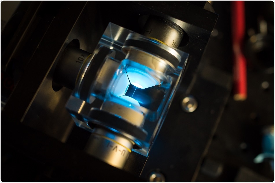
Hari Shroff, from the National Institute of Biomedical Imaging and Bioengineering (NIBIB), stated that “in one sense, it [the technique] couldn't be easier”. Commercially available mirrored cover slips were utilized instead of glass cover slips to grow the biological samples.
Then the samples were mounted on the dual-view inverted selective plane illumination microscope (diSPIM) invented in Shroff's laboratory, which is comprised of two objectives delivering perpendicular views of the sample.
In the usual diSPIM setup, a thin sheet of light is transmitted to the sample by one objective while the image is collected by a camera positioned behind the second objective. Then the functions of objectives are alternated as follows: a thin section of the sample is illuminated by one objective in a perpendicular direction while the other objective images it.
Alternatively, with mirrored cover slips, the transmitted light along with the fluorescence produced by it in the sample reflects off the mirror, so that four complementary views of the sample can be simultaneously collected by the two objectives.
This significantly enhances the light collection efficiency of the microscope and doubles its speed, which is beneficial for imaging fast-moving biological samples and processes with low light.
Achieving high speed while collecting more information is only half the battle: computational resolution of the technique is required to produce an image. Patrick La Rivière, from the University of Chicago, led the team in developing an algorithm to optimize spatial resolution and fuse the views in all three dimensions (x, y, and z).
La Rivière suggested that the computation is really enabling for this technique. He further added: "While the mirror multiplies the information captured by the cameras, it also introduces some contamination that would not normally be there. What we were able to show is by properly modeling the process - basically, by converting the microscope into mathematics - we could effectively remove that contamination and recover all the information (imaged by the cameras)."
The applicability of the technique was demonstrated by the team by imaging a variety of live samples, such as calcium activity in nematode embryos and Golgi, microtubule, membrane, and mitochondrial dynamics in cells.
 AI-guided microneedle patch changes shape to heal chronic wounds