Oct 19 2018
Human neurons are much larger than those of model organisms mice and rats, so it's been unclear whether it's size that makes a difference in our brain's computational power. Now, in a study appearing October 18 in the journal Cell, researchers show that unlike those of other animals, human neurons employ highly compartmentalized signaling. Human dendrites--the tree-like branching structures that function as neurons' antennas--process electrical signals differently than dendrites in rodents, the most common model systems for studying neuronal properties.
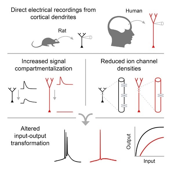
"The human neuron is basically like a rat neuron, but because it's so much longer, signals have much farther to travel. The human dendrites thus have a different input-output function" from rats, says senior author Mark Harnett, the Fred and Carole Middleton Career Development Assistant Professor in the Department of Brain and Cognitive Sciences at the Massachusetts Institute of Technology. "Dendrites farther away from the cell body have fewer ion channels, which control signal processing. That was something we absolutely did not expect."
Harnett, who studies how the biophysical features of neurons shape information processing in the brain, believes our longer, bigger dendritic arbors endow human neurons and their respective circuits with enhanced computational abilities.
"Human neurons are more compartmentalized electrically and can exploit this," he says. "We think that having low ion channel density at the ends of dendrites lets the cell have as many subcompartments as possible. The longer the branches, the more independent the units. You have many more units to do computation within a single neuron."
"Integrating different streams of information in this manner could endow individual neurons with the sophistication of small computational networks," says Harnett.
Using a technique called patch-clamp recording, in which tiny glass needles are sealed against the cell membrane to measure detailed electrical properties, the researchers for the first time directly recorded dendritic activity in living brain tissue from humans. The human tissue (from brain surgeries) was obtained from the anterior temporal lobe of epilepsy patients.
The work could also eventually benefit patients with epilepsy, in which small sections of brain tissue are sometimes removed to control seizures that don't respond to medication. "People have used animal models to think about epilepsy for a long time, but clearly, there are some pretty significant differences, at least in the dendrites, between humans and rodents," Harnett says. "The better we understand ion channels and membrane excitability, the more insight we gain into the mechanisms of epilepsy and how to treat it."
Next steps also involve determining the relationship between neuron size and electrical properties in other species to gain insight into the evolution of the cortex.
Source: https://www.cell.com/
 Western diet weakens the gut’s nervous system through iron-dependent damage