Cardiovascular disease (CVD) is the leading cause of death worldwide, with over 17 million per year according to the World Health Organization. Many forms of CVD are chronic in nature, meaning that they worsen over time. Thus, once the disease has been diagnosed it is important to initiate treatment as soon as possible in order to provide positive patient outcomes.

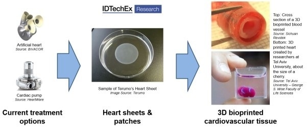
Moving towards 3D bioprinting cardiovascular tissue. Source: IDTechEx Report “Cardiovascular Disease 2020-2030”” (www.IDTechEx.com/CVD)
The ideal treatment for some forms of severe CVD, such as chronic heart failure or extensive myocardial injury, is cardiac transplantation. Due to shortages in available donor tissue, this cannot be given to all patients. The average waiting time for a suitable donor is six to twelve months in the USA and around one in six people die before they can receive a transplant. There is a clear need for a more abundant supply of hearts suitable for transplantation.
Until significant progress has been made to boost this supply, cardiologists need to rely on the technology at their disposal. A common strategy to address heart failure is to use a cardiac pump such as a left ventricular assist device (LVAD). Mechanized hearts have also been explored as a treatment option for chronic heart failure when transplant donors are not available.
Current treatment options are useful to a certain extent, but personalized solutions are required to improve patient outcomes and quality of life. This need is driving the development of cardiovascular 3D bioprinting technologies, which make use of 3D printing-like techniques to combine cells and biomaterials to fabricate biomimetic structures that replicate natural tissue physiology and function.
Developing a dynamic cardiac tissue capable of mimicking the mechanical and electroconductive properties of native myocardium is proving difficult for researchers. Many challenges stand in their way including, among others, re-creating tissue matrix and providing an adequate oxygen supply to each cell.
The success of 3D bioprinting depends on researchers’ ability to vascularise the tissue. For this reason, a lot of focus has recently been placed on the generation of blood vessels. Several promising studies have already been conducted. For instance, researchers at University of California San Diego 3D printed a functional blood vessel network which, once implanted in mice, merged with the animal’s blood vessels and was capable of transporting blood. Similar achievements have been reported by Sichuan Revotek, Rice University and the University of Pennsylvania in the last few years.
An important innovation as we move towards 3D bioprinting cardiac tissue is the development of cell sheets. Terumo, a Japanese conglomerate, has commercialized the Heart Sheet for treatment of heart failure in Japan. To develop Heart Sheet, muscle tissue is harvested from the patient’s leg and cultured in vitro. Terumo has developed a tissue culture plate that allows cells to float off the surface in an intact sheet when the temperature is lowered, thus preserving the extracellular matrix that is lost when cells are removed by other methods.
Cardiac tissue engineering techniques such as this one can be used to create functional constructs capable of re-establishing the structure and function of damaged myocardium following myocardial infarction. The engineered cardiac tissue, which often comes in the form of a “patch”, is implanted directly onto scar tissue. The intention is to compensate for the heart’s reduced function by strengthening its structure and boosting its ability to pump blood. This way, researchers hope to reduce the need for transplants, improve recovery and prevent subsequent events.
Researchers across the world are developing “cardiac patches”. In June 2019, Imperial College London announced the creation of thumb-size patches of heart tissue that start to beat spontaneously after three days and start to mimic mature heart tissue within one month. These patches successfully led to improvements in heart function following a heart attack after only four weeks. Importantly, blood vessels appeared to have formed within the patch after that time. Clinical trials are expected to be held in 2020 or 2021.
Once implanted, cardiac patches could do more than just promote cardiac tissue regeneration. For instance, a bionic patch could deliver electrical shocks and act as a pacemaker. Scientists at the University of Tel Aviv also investigated integrating electronic sensors into the patch to enable remote monitoring of cardiac activity.
Although researchers have not yet been able to create a fully functioning artificial heart, an important leap was made in 2019. Researchers from Tel Aviv University unveiled the first 3D bioprinted heart with human tissue including chambers, ventricles and blood vessels. To accomplish this, a biopsy of fatty tissue from patients was taken to produce the cells required. Patient-specific cardiac patches were produced first, after which the entire heart was made. Although the heart is capable of contracting, it remains a long way off from being ready for clinical trials as it cannot yet pump blood and is the size of a cherry.
3D bioprinting has the potential to provide a heart or blood vessels to patients in need of transplants. The tissue would be made from their own cells, thereby considerably reducing the risk of rejection. Despite promising recent innovations, 3D bioprinting technology remains in its early days and is unlikely to become a viable therapeutic option in the near future due to the numerous roadblocks (both technical and regulatory) that it currently faces. This will change once the technology evolves and full-sized hearts and vessels can be constructed efficiently and at scale.
To find out more about technologies for 3D bioprinting cardiovascular tissue, or on the topic of cardiovascular disease management as a whole, please refer to IDTechEx’s report: “Cardiovascular Disease 2020-2030” available on the IDTechEx website at www.IDTechEx.com/CVD.
To connect with others on this topic, join us at The IDTechEx Show! USA 2020, November 18-19 2020, Santa Clara, USA. Presenting the latest emerging technologies at one event, with six concurrent conferences and a single exhibition covering 3D Printing and 3D Electronics, Electric Vehicles, Energy Storage, Graphene & 2D Materials, Healthcare, Internet of Things, Printed Electronics, Sensors and Wearable Technology. Please visit www.IDTechEx.com/USA to find out more.
IDTechEx guides your strategic business decisions through its Research, Consultancy and Event products, helping you profit from emerging technologies. For more information on IDTechEx Research and Consultancy contact [email protected] or visit www.IDTechEx.com.