Sponsored Content by TissueGnosticsReviewed by Maria OsipovaJul 1 2025
In this interview, News Med talks to Prof. Uwe Ritter about the role of advanced imaging techniques in uncovering the role of CD8+ T cells in tissue regeneration.
What are the main goals of your research into immune cell interactions and tissue regeneration?
My work focuses on exploring immune cell interactions and tissue regeneration through cutting-edge imaging technologies, most notably the image processing and analysis pipelines of 3D skin models. These models are beneficial for characterizing regular T cells (Tregs) in wound healing.
I am also using machine learning to analyze organoid cultures, particularly focused on investigating the impact of CD8+ T cells on tissue remodeling and the decryption of cell-cell interactions in situ.
Mouse experiments involving the depletion of Tregs (pan depletion, subset-specific depletion, or induced depletion) have shown that Tregs are involved in immune regulation, metabolism, and tissue repair. These Tregs are characterized by the expression of amphiregulin and Foxp3.
The repair function in murine systems is well established, but we would like to know whether or not there are equivalent tissue repair Tregs in humans.
Michael Sforer's group analyzed mouse and human tissues to investigate this equivalence. They analyzed blood, subcutaneous fat, and skin, performing single-cell attacks to look for any overlap in markers expressed by human or mouse Tregs to detect the phenotype of tissue repair Tregs in humans.
This research revealed that it was possible to detect tissue Tregs in humans that express CCR8 and are positive for basic leucine zipper ATF-like transcription factor (BATF). These are comparable to tissue Tregs from the mouse immune system.
Could you explain how imaging pipelines support the investigation of tissue regeneration processes?
In the example I discussed, molecular analysis revealed that these tissue Tregs included Tfh, a molecular helper T cell differentiation program. This discovery offers a major advantage because we can generate Tfh-like tissue Tregs and naive T cells and look for functional analysis or tissue repair functions.
The pipeline for this experiment involved isolating the tissue Tregs from PBMCs from blood and enriching them with CD25. We performed FACS sorting for the markers CD45 and CD45RA. We isolated antigen-naive Tregs and stimulated those naive Tregs with IL-2 to generate IL-2-derived Tregs. This allowed us to use Tfh-like Tregs similar to tissue Tregs, adding components such as IL-2, IL-12, IL-21, IL-23, and TGF-b.
We added TransAct IL-2 to these T cells, allowing us to further activate and harvest the supernatant from these Tregs. This process gave us two supernatants from the IL-2 Tregs and putative repair Tregs from the tissue itself.
What makes 3D skin models particularly useful for studying Tregs in wound healing? Could you expand on your work in this area?
We contacted Florian Gruber in Vienna and Dirk Becker in Würzburg because this group had established a very useful 3D skin model involving growing human epidermal keratinocytes in plastic (PET) tubes.
As the keratinocytes grow, they build up a strong epidermal compartment. A dermal punch can then be used to punch a hole into the epidermal compartment, and the keratinocytes start to close this area. This is essentially wound healing.
We aimed to determine whether supernatants from Tfh-like Tregs or IL-2 Tregs better accelerate wound healing. Using a time kinetic approach, we cut the epidermal pieces in a cryostat.
We aimed to measure the newly built stratum corneum in this structure. We needed to develop a software-based solution for dissecting structures from this epidermal compartment because we wanted to quantify the new stratum corneum in relation to the root healing effect of supernatant from the different Tregs I mentioned.
The challenge was achieving the sophisticated color and structural separation to properly discern the epidermal tissue, the stratum granulosum, the stratum basale, and the polycarbonate matrix of the 3D skin model.
The software detected the structures present in the epidermal compartment, reducing the impact of the background by subtracting the shades that do not correlate with the epidermal compartment. This is what allowed the stratum corneum to be detected.
The software can be used to measure the number of keratinocytes in the epidermal compartment, and it is possible to use advanced color separation using special settings for tissue detection, polycarbonate detection, and more.
The software’s capabilities were key to answering whether the supernatant could increase the wound healing in the system. The results showed that the IL-2 Treg supernatants did not have a significant impact on wound healing, but the Tfh-like cells supernatant saw the square micrometer of the stratum corneum become normalized.
This technique can be reproduced via other methods such as trans-epithelial electrical resistance (TEER) measurements. Comparing these measurements showed similarity between the different approaches, confirming that the technique was working.
We developed a detection algorithm that could dissect the pink shades of tissue. We also showed that tissue Tregs from humans are CCR8- and BAFT-positive and that these Tregs could accelerate wound healing in more complex wound-healing systems.
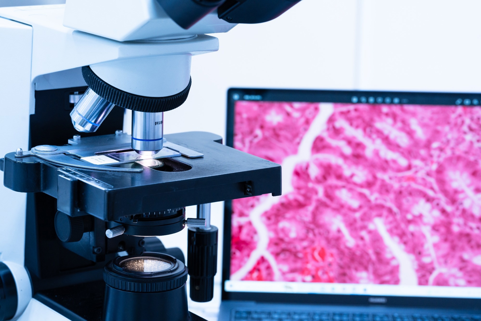
Image Credit: Komsan Loonprom/Shutterstock.com
How are you using machine learning with organoid cultures to study CD8+ T cells?
CD8+ T cells are well known for their ability to kill virus-infected cells or tumor cells. CD8+ T cells in the dysfunctional state (called exhaustion) are positive for PD1, TIGIT, and other markers.
Immunity is not black and white, however. In systems like liver hepatocellular carcinoma or NASH, it has been shown that CD8+ T cells that are PD1+ and TIGIT+ are reactive. They are not exhausted and can promote fibrosis or tumor progression.
We want to know whether these CD8+ T cells are involved in tissue reconstruction. When CD8+ T cells kill infected cells, we see tissue destruction that requires remodeling. Therefore, we can ask whether CD8+ T cells can also induce tissue regeneration.
To investigate this, we isolated those T cells and stimulated those CD8+ T cells using CD3 and CD28 beads. We took the resulting supernatant and performed an easy scratch assay. Activated naive T cells are not useful in wound healing, but we did observe that activated PD1+, TIGIT+, and activated central memory T cells can induce wound healing.
We also wanted to know whether CD8+ cells can promote organoids' reorganization. To investigate this, we isolated CD8+ T cells using the TexMACS medium, activated those T cells (because only activated CD8+ T cells can induce wound healing), and added them to organoids.
Organoids are simple tissue-engineered cell-based in vitro models that recapitulate many aspects of the complex structures and functions of corresponding in vivo tissues. Organoids are tissue-derived and we can derive them from adult stem cells.
Cancer stem cells are induced through pretense stem cells, so this is not a problem. We can generate organoids in a Matrigel dome, so we opted to use this approach.
For example, we wanted to know what happens when the organoids come into close contact with lymphocytes. This had not been done before our study because measuring and dissecting organoids from lymphoids is very difficult.
With this in mind, our goal was to develop a machine learning pipeline suitable for detecting organ structures within the complex co-culture and high-throughput conditions. This had to include the characterization of organ formation, including numbers, size, and shape.
We began by deriving organoids from the bile duct and combining them with CD8+ T cells. Then, we started working in collaboration with TissueGnostics to develop an organoid detection app.
A number of problems had to be addressed at the beginning of this process. For example, we saw contour mimicry whereby cells look like organoids but are not organoids. We also saw contour fusion, with a fusion of two organoids that are difficult to dissect, and contour disruption, which makes it difficult for the algorithm to detect the organoid structure.
The software takes the organoid image, converts it to grayscale, and performs a background correction. The software then allows one to see the membrane of the organoids, distinguishing between the membrane and the organoids and the background of the lymphocytes and other structures. Corrections can be applied as required to better split the background from the organoids and implement a layer specification of the organoids.
It is also possible to adjust size and compactness when you are not confident or happy with the results.
We used the classifier machine learning system to improve this new system. For example, it is possible to train the algorithm by manually marking the tissue and marking the organoid boundary.
This type of manual delineation is time-consuming, but it was a useful quality control measure. We used a linear regression model to compare organoid size using both manual detection and software detection, confirming that the app is very precise and that it can be used to perform studies.
Results showed an increase in CD8+ cells from different donors. We measured the total area and sum of the organoids, showing an increase in organoid development in the presence of CD8+ T cells.
Our findings also indicate that the cytotoxic effector program of CD8+ cells is closely linked to a regeneration program, both of which are evoked upon a teaser stimulation. CD8+ T cells can kill other cells, but they also induce a tissue regeneration program.
What are your visions for decoding cell-cell interactions in situ using advanced imaging technologies?
I think we will be able to effectively decode cell-cell interactions in a few years. Cell communication between immunological cells such as T cells or dendritic cells is often based on autocrine signaling or juxtaposition processes. These processes influence tissue homeostasis and immune response to tumors and pathogens.
There are signaling molecules involved, for example, cytokines, metabolites, receptor proteins that bind specific surface molecules, signaling pairs, cell junctions, and secondary messengers. In terms of understanding this communication, we know the alphabet, but we do not currently understand the sentences.
Many people are already working with single-cell RNA-seqs and single-cell FACS analysis, allowing them to describe the transcriptome and phenotype of each single cell. However, we still do not know which cell has been in contact with other cells or which cell is responsible for activating other cells. These questions cannot be answered via single-cell analysis.
We can generate single-cell raw data at the transcriptome, proteome, or metabolome level, but this is the limit at the moment.
We can perform very simple in vitro experiments. For example, we can evaluate OT-1 T cells specific for ovalbumin by adding ovalbumin to an in vitro culture with dendritic cells and measuring the immune response of the dendritic and T cells.
We can also use laser scanning microscopy to measure the membrane overlaps of cells (immunological synapse) to characterize the phenotype of cells that interact with dendritic cells. This approach is very precise, but it only describes the phenotypes.
We can also do this in situ, measuring cell boundaries via the StrataQuest software. This allows us to calculate the potential contact area of cells, analyzing the cells that are in direct contact with the analyzed cell to characterize its neighbors. However, this method does not show whether cells are in communication.
We have performed experiments. We took APCs, defined endogen, a T cell receptor, and added ovalbumin. We then measured single-cell and double-cell effects with RNAseq and dcRNAseq. After sorting the droplets, we analyzed the phenotype modification or the transcriptome of those cells interacting in terms of their phenotype modification or non-phenotype modification.
How does your work in this area correlate with the PIC-seq methods developed by Ido Amit’s lab?
It is closely aligned. In 2020, an excellent publication by Ido Amit described a similar approach to ours. They did exactly the same. They kept it simple, used APCs, defined a TCR receptor, isolated the T cells and dendritic cells, and then conjugated doublets. They then performed single-cell analysis and physically interacting cells (PIC) analysis.
This allowed them to develop a PIC-seq algorithm that enabled the dissection of dendritic cells and T cells. This PIC-seq approach allowed the team to analyze the core signature of the attached dendritic cells or T cells.
This is very important because this approach removes the need to isolate and dissect doublets, allowing us to begin to characterize the crosstalk of physically interacting cells.
However, it is important to note that this approach is only possible when the two cell subsets are transcriptionally distant. When they are transcriptionally related, this technique is not functional.
I became fascinated with this technique and wrote an opinion article about it that was published this year. I explained that it is possible to analyze single cells, analyze PICs, sequence single cells on the PICs, and run the PIC-seq. This is based on the pre-processing, integration, deconvolution, and differentiation of the data.
My idea was that it is also possible to define the gene expression profiles of PICs grouped by the contributing T cell or myeloid identity. For example, it would be possible to characterize migratory DCs based on their genes, plasmacytoid DCs, DC-1, DC-2, or monocytes. A similar definition is possible with T cells, for example, characterizing a naive T cell, CD4+ Treg, CD8+ CTL, or an activated T cell.
When we know the profile of physically interacting cells based on experimental data with ovalbumin or other antigens, we can determine the gene profiles of migratory antigen-presenting TCs and know exactly what they are producing.
This allows us to create a panel, choosing antibodies with fluorescence, for example, and performing multiplex staining.
This requires a microscope capable of imaging multiple channels. The key advantage of this approach is that we can detect the cells that are genuinely interacting based on the presence of specific markers.
What types of research do you see potentially evolving from these approaches?
This approach is potentially valuable for mouse systems where, for example, we can infect mice with pathogens, and because we know we have a resistant phenotype and a chronic phenotype, we can detect the gene modules that correlate to adaptive immunity by T cells or dendritic cells that correlate to immune pathology.
There are differences between viruses, protozoans, and bacteria, but this approach could lead to the creation of a multicenter-based contextual and PIC atlas, featuring relevant gene modules and a pathogen-specific background.
For example, during the immune response of the PICs, we may see shared expressions of genes also induced by protozoans or bacteria. The software could be used to detect genes that are exclusively induced by virus infection. We could isolate these genes using future software packages, demonstrate these findings, and relate these back to the in situ situation, clearly showing or describing the cells in communication in a pathogen-specific manner.
These findings could also eliminate the need to work with dendritic and T-cells. We may see this concept expand into other systems, such as tumor immunology. It may become possible to characterize gene models associated with tumor-killing based on the interaction between cells and the expression of genes that are specifically induced during tumor-killing processes.
This type of computational analysis will allow us to pinpoint biomarkers, for example, that are associated with tumor immune cell interaction. The advantages of these systems are clear: they allow us to deepen our understanding of tumor immune communications, identify tumor gene modules, and characterize tumor escape mechanisms.
I also think this will improve the interpretation of tumor-effector cell interactions in situ, which will be crucial for evaluating tumor progression and treatment activities.
About Prof. Uwe Ritter
Prof. Uwe Ritter is a principal investigator in the Department of Immunology, Leibniz institute for Immunotherapy. His research is focused on studying the immune system's response to various antigens in the skin-associated lymphatic tissue (SALT). He and his team aim to manipulate critical immune checkpoints in myeloid cells to potentially reprogram dysfunctional immune reactions. Their work integrates cutting-edge technologies – notably in-situ contextual tissue cytometry and high-resolution imaging – to gain deeper insights into tissue structure and function.
About TissueGnostics
TissueGnostics (TG) is an Austrian company focusing on integrated solutions for high content and/or high throughput scanning and analysis of biomedical, veterinary, natural sciences, and technical microscopy samples.
TG has been founded by scientists from the Vienna University Hospital (AKH) in 2003. It is now a globally active company with subsidiaries in the EU, the USA, and China, and customers in 30 countries.
TissueGnostics portfolio
TG scanning systems are currently based on versatile automated microscopy systems with or without image analysis capabilities. We strive to provide cutting-edge technology solutions, such as multispectral imaging and context-based image analysis as well as established features like Z-Stacking and Extended Focus. This is combined with a strong emphasis on automation, ease of use of all solutions, and the production of publication-ready data.
The TG systems offer integrated workflows, i.e. scan and analysis, for digital slides or images of tissue sections, Tissue Microarrays (TMA), cell culture monolayers, smears, and other samples on slides and oversized slides, in Microtiter plates, Petri dishes and specialized sample containers. TG also provides dedicated workflows for FISH, CISH, and other dot structures.
TG analysis software apart from being integrated into full systems is fully standalone capable and supports a wide variety of scanner image formats as well as digital images taken with any microscope.
TG cooperations
TG continuously cooperates with research groups and other companies in the industry to provide novel tools and applications to its customers.
 Exploring the role of immune checkpoints IDO1 and PDL1 in Cutaneous Leishmaniasis (CL)