Sponsored Content by TissueGnosticsReviewed by Maria OsipovaOct 24 2022
Colorectal cancer is the second most prevalent cause of cancer-related mortality.1 An important contributing factor to this statistic is the high risk of liver metastases. Liver metastases can affect up to half of colorectal cancer patients as treatment options are limited.2
Liver surgery is the main type of treatment for patients with colorectal liver metastases (CRLMs); however, this is not practical for all patients. As a result, alternative treatment options are being sought. Targetting the anti- or pro-tumoral immune response is a new approach that is being taken in cancer treatment.
Tumor-infiltrating immune cells include cellular populations with a wide range of functions and phenotypes.3 For example, the distribution and profile of intra-tumoral T cells (TILs) are key considerations when evaluating prognosis in primary colorectal cancer.4 Additionally, evidence indicates that a positive prognosis in CRLM correlates with CD8+TIL levels in CRLM.5,6
In terms of clinical prognosis, additional immune cell types, such as B cells, play a critical role as they form B-cell-enriched and highly proliferative immune structures in the vicinity of cancer tissue, which are shown to be prognostic towards an improved clinical outcome.7
Regarding T cells, high concentrations of intra-tumoral CD8+ tissue-resident memory T cells (TRM cells) have been associated with increased survival in primary colorectal cancer.8 With more studies demonstrating the significance of TILs in anti-tumor responses, TILs may become a possible target for new treatment approaches in CRLM.
To develop these alternative therapeutic options, the characterization of tumor-reactive CD8+ TILs is necessary. In particular, it would be valuable if the significant differences in TIL composition between healthy liver and CRLM could be identified.
Some studies have reported that higher concentrations of CD103+CD39+CD8+ TRM cells occur in CRLM relative to adjacent liver tissue.9 The specific CD8 cell subpopulations have since been evaluated in detail, which was facilitated by the TissueFAXS CHROMA imaging platform10 and StrataQuest image analysis software.11
Evaluating CD8 cells in colorectal liver metastases
Recent research using TissueFAXS CHROMA and StrataQuest assessed the activation states and frequencies of CD103+CD8+ TRM cells and other T cell populations in CRLM, as well as adjacent liver tissue in samples from patients with CRC.
Another aim of this research was to determine whether CD8+ TRM cells accrued in subcutaneous CRLM-derived tumors in lymphocyte-deficient mice.12
Using the automated scanning function of TissueFAXS CHROMA,10 the samples were imaged as entire tissue at 20x magnification. The images acquired by TissueFAXS CHROMA were then analyzed using the StrataQuest software.11 DAPI staining was used to detect individual cells, whilst specific marker staining was used to classify specific cell phenotypes.
By using a machine-learning process, the StrataQuest software allowed tumor cells and stromal cells to be identified and separated. Additionally, StrataQuest was able to group stromal cells based on their distance from the tumor.
Findings
Together, the TissueFAXS CHROMA and StrataQuest technologies have enabled the detection of distinct immune microenvironments of CRLM and the adjacent liver. CRLM tissue samples revealed decreased levels of CD8+ αβ T cells and increased frequencies of CD4+ αβ T cells.12
By studying the capability of the predominant TILs to control CRLM-derived tumor growth in vivo using a patient-derived xenograft model, researchers have discovered that the transferred TILs only succeeded in controlling the growth of CRLM-derived tumors after trametinib treatment.
Intriguingly, CD103+CD8+ TRM cells only accumulated within the autologous CRLM-derived tumor rather than in the spleen or blood.
The susceptibility of CD103+CD8+ TRM cells for the repopulation of the autologous tumor and the occurrence of CD103+CD39+CD8+ TRM cells in CRLM adjacent to the liver suggests that further investigation into the viability of CD103+CD39+CD8+ TRM as potential targets for treatments against advanced CRLM is required.
Summary
CD103+CD39+CD8+ TRM cells accrued in CRLM relative to adjacent liver tissue, emphasizing that they are potential targets for the design of novel treatment approaches for CRLM.
These functional and phenotypic analyses of TILs in CRLM using TissueFAXS CHROMA and StrataQuest may assist the development of novel TIL-mediated treatments for colorectal cancer and liver metastases.
The new data acquired using TissueFAXS CHROMA and StrataQuest is critical as it highlights their potential application in research, such as tackling the treatment of advanced colorectal cancer in patients with inoperable liver metastases.
TissueFAXS CHROMA – Multipectal high-speed imaging
The multipurpose TissueFAXS CHROMA is a multifluorescence imaging platform that scans the whole slides concurrently for up to 7 markers while preventing any bleed-through. The sophisticated image analysis solution, StrataQuest, comes with TissueFAXS CHROMA and therefore provides a holistic solution for in-depth tissue microenvironment characterization and cellular phenotyping.
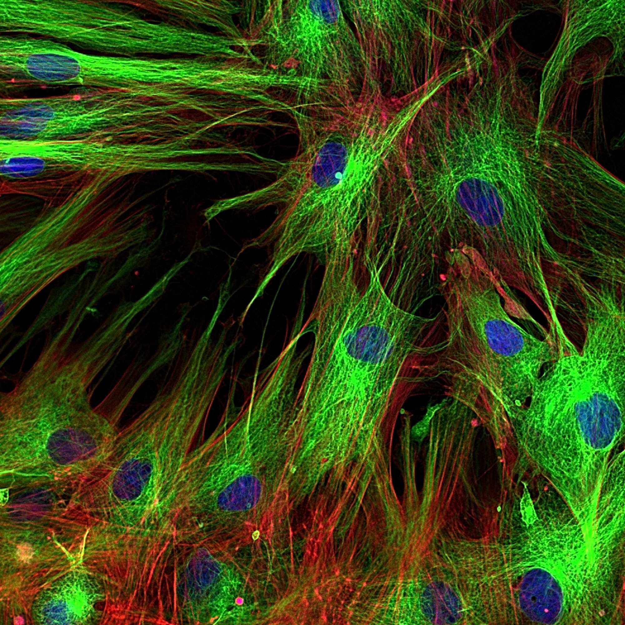
Image Credit: Vshkova/ Shutterstock
StrataQuest
An array of streamlined analysis solutions, including the sophisticated cell-to-cell Contact App, are provided by the StrataQuest software.11 The Contact App facilitates the identification of the cellular phenotype of specific fluorescence-stained cell populations and forms cellular contacts with their neighboring cells.
It readily delivers the staining intensity per marker and morphometric parameters for each segmented cell/cell compartment. Additionally, StrataQuest makes it possible to detect cells in situ that are defined by more than one marker. This formed a critical part of this research by detecting CD3+, CD103+, CD8+, and TRM.
The StrataQuest analysis is conducted using a Cell-to-Cell Contact App that is available online in the TissueGnostics App Center.
References
- Sung H, et al. Global Cancer Statistics 2020: GLOBOCAN Estimates of Incidence and Mortality Worldwide for 36 Cancers in 185 Countries. CA Cancer J. Clin. 2021, 71, 209–249.
- Martin J, et al. Colorectal liver metastases: Current management and future perspectives. World J. Clin. Oncol. 2020, 11, 761–808.
- Bartsch LM, et al. Tissue-Resident Memory T Cells in the Liver-Unique Characteristics of Local Specialists. Cells 2020, 9, 2457.
- Pages F, et al. In situ cytotoxic and memory T cells predict outcome in patients with early-stage colorectal cancer. J. Clin. Oncol. 2009,27, 5944–5951.
- Liang JY, et al. Histopathological growth patterns correlate with the immunoscore in colorectal cancer liver metastasis patients after hepatectomy. Cancer Immunol. Immunother. 2020, 69, 2623–2634.
- Wang E, et al. Prognostic value of the density of tumor-infiltrating lymphocytes in colorectal cancer liver metastases. Oncol. Lett. 2021, 22, 837.
- Mungenast, F. et al. The Immune Phenotype of Isolated Lymphoid Structures in Non-Tumorous Colon Mucosa Encrypts the Information on Pathobiology of Metastatic Colorectal Cancer. Cancers 2020, 12, 3117. https://doi.org/10.3390/cancers12113117
- Okla K, et al, Tissue-resident memory T cells in tumor immunity and immunotherapy. J. Exp. Med. 2021,218, e20201605.
- Liang F, et al. Antigen Presenting Cells from Tumor and Colon of Colorectal Cancer Patients Are Distinct in Activation and Functional Status, but Comparably Responsive to Activated T Cells. Cancers 2021, 13, 5247.
- TissueGnostics.
- TissueGnostics
- Liang F, et al. A Fraction of CD8+ T Cells from Colorectal Liver Metastases Preferentially Repopulate Autologous Patient-Derived Xenograft Tumors as Tissue-Resident Memory T Cells. Cancers 2022, 14,2882.
About TissueGnostics
TissueGnostics (TG) is an Austrian company focusing on integrated solutions for high content and/or high throughput scanning and analysis of biomedical, veterinary, natural sciences and technical microscopy samples.
TG was founded by scientists from the Vienna University Hospital (AKH) in 2003. It is now a globally active company with subsidiaries in the EU, the USA and China and customers in 28 countries.
TG systems offer integrated workflows, i.e. scan and analysis, for digital slides or images of tissue sections, Tissue Microarrays (TMA), cell culture monolayers, smears and of other samples on slides and oversized slides, in Microtiter plates, Petri dishes and specialized sample containers.
Sponsored Content Policy: News-Medical.net publishes articles and related content that may be derived from sources where we have existing commercial relationships, provided such content adds value to the core editorial ethos of News-Medical.Net which is to educate and inform site visitors interested in medical research, science, medical devices and treatments.