Mid-infrared (Mid-IR) spectroscopic imaging, alternatively referred to as chemical imaging, is a powerful label-free, non-destructive, quantitative technique that allows a user to see both the chemical and morphological structure of a heterogeneous sample-; a single, rapid measurement can reveal the presence of chemical species, their quantities and their precise locations without stains, dyes, or fluorescent tags.
Mid-IR chemical imaging has been widely applied in in a range of research and industrial applications over the past two decades to great success. However, it is only relatively recently with the advent of new technology, that the technique has produced chemical images precise enough and fast enough for the robust study of biological tissue1.
Spero Advantages
Chemical imaging in disease diagnosis
In traditional histopathology, tissue samples are stained in order to identify different cell types or to label specific biomolecules for diagnostic purposes. However, the staining process is destructive, fundamentally altering the chemical structure of the specimen and this prevents the tissue section from being analyzed by follow-on molecular analyses. This is of particular importance in pre-clinical drug development where subjecting a single tissue section to a multitude of analyses is essential. Furthermore, in many cases such as during surgery, staining a biopsied section followed by a visual inspection under a traditional microscope by a trained pathologist can be time-prohibitive.
Mid-IR chemical imaging takes advantage of the fact that different biomolecules in a sample absorb portions of the infrared spectrum differently. Healthy and diseased tissues exhibit distinct chemical signatures. Therefore, it is possible to spatially differentiate between diseased and healthy tissue without applying any stains. This leaves the specimen chemically and structurally intact for downstream genomic or proteomic analyses. Furthermore, mid-IR chemical imaging has been used to distinguish grade and sub-typing of disease. Consequently, a sample can be prepared and evaluated more quickly, allowing a diagnosis to be reached more rapidly for example in an operating theater.
The analysis can also be automated using readily-available chemometric analysis software to assist the pathologist in making a diagnosis 2. Furthermore, the technique provides additional information about the metabolic state of cells, which may not be accessible using conventional staining techniques.
The interpretation of stained images of liver biopsies remains a challenge to pathologists when diagnosing the presence and extent of liver disease. The difference between healthy liver cells and scarring—fibrosis—as a result of repeated damage by disease, e.g., hepatitis, toxins, or alcohol, is not always obvious during imaging. Yet this is an important distinction to make since fibrosis can ultimately lead to a loss of liver function, known as cirrhosis, and even cancer. Cirrhosis can arise by many different routes and so is associated with many different chemical signatures, making it difficult to determine the severity of the damage and assess the likely prognosis.
Comorbid conditions such as diabetes can further complicate the chemical signatures obtained from liver biopsies. There are consequently a host of variables that must be considered when deriving useful diagnostic or prognostic information from chemical imaging of liver biopsies. It is hoped that recent advancements in quantum cascade laser based infrared spectroscopic imaging may help.
Quantum cascade laser infrared spectroscopic imaging
Quantum cascade laser infrared spectroscopic imaging uses a broadly tunable quantum cascade laser(QCL) as the light source allowing for high-SNR discrete-frequency imaging at video rates3.
The ability to specifically target a particular spectral frequency means the imaging can be tuned to the tissue features of interest so they can be more readily identified in real-time. This is of particular interest when trying to reach a diagnosis from a liver biopsy since it makes it possible to differentiate the different structural features of the liver more easily.
Furthermore, the ability to select the frequencies at which a sample is analyzed enables the acquisition of manageable datasets from spectroscopic imaging of large samples. Previously, the size of the dataset obtained from spectroscopy of a large sample was too cumbersome to make analysis feasible. The image of a 192 mm2 colon tissue section obtained using QCL infrared spectroscopy comprised 11 million pixel spectra and was less than 1GB in size3.
Recent research using this novel technique was able to discriminate between healthy hepatocytes and fibrotic tissue in liver biopsies4. It also demonstrated how the chemical signatures of these cells are modified according to the diabetic status of the patient4. Furthermore, QCL-infrared spectroscopic imaging identified specific chemical signatures relating to different severities of liver disease. This novel technique could be considered a breakthrough in liver disease diagnostics and research.
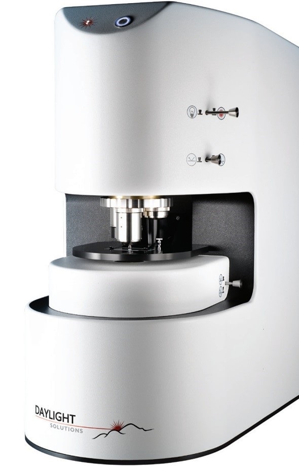
The Spero® QCL imaging system
Spero® (Daylight Solutions) is the world's first and only commercially available laser-based infrared microscope. The advanced QCL-infrared spectroscopy technology is incorporated into a small desktop microscope5. The ultra-bright, tunable laser light source provides simultaneous high-resolution with a wide field of view. Together, these features allow a rapid through-put of samples that can speed up sample screening by orders of magnitude. In addition, Spero has a live mode that enables imaging to be obtained in real-time. In full data collection mode, a complete spectral scan can be collected in seconds.
Summary
QCL‐based infrared spectroscopy has expanded the potential of imaging in pathological analysis, allowing greater detail to be obtained in the fraction of the time of conventional infrared spectroscopy. In such a dynamic field, this novel imaging technology will undoubtedly become invaluable in a wide range of applications.
References and Further Reading
- Lyra da Cunha MM, Trepout S, Messaoudi C, et al. Overview of chemical imaging methods to address biological questions. Micron 2016;84:23–36.
- Fernandez DC, Bhargava R, Hewitt SM, et al. Infrared spectroscopic imaging for histopathologic recognition. Nature Biotechnology 2005;23:469‑474
- Daylight Solutions Application Note 201602. Available at https://www.daylightsolutions.com/examining-tissue-pathology-using-quantum-cascade-laser-qcl-mid-infrared-spectroscopic-imaging/
- Sreedhar H, Varma VK, Gambacorta FV, et al. Infrared spectroscopic imaging detects chemical modifications in liver fibrosis due to diabetes and disease. Biomed Opt Express 2016; 7(6):2419‑2424.
- Daylight Solutions website. http://www.daylightsolutions.com/life-sciences/spero.htm. Accessed 29 November 2016

About Daylight Solutions
Daylight Solutions’ molecular detection and imaging products consist primarily of lasers, sensors, and imaging systems, all of which leverage the company’s mid-infrared, quantum cascade laser (QCL) technology. This core technology provides a versatile platform from which new products are developed, allowing the company to serve markets that include Scientific Research, Life Sciences, Defense, and Commercial.
The company is committed to innovation and introduced the world’s first broadly tunable mid-infrared laser system for scientific research, the world’s first semiconductor-based laser for protecting aircraft against shoulder-fired missiles, and the world’s first mid-infrared laser-based microscope for real-time biochemical imaging and material analysis.
Daylight Solutions consists of two separate business units. In 2009 the company created a wholly owned subsidiary to address the specific requirements of the defense industry. As a subsidiary, Daylight Defense developed the business and manufacturing infrastructure necessary to deliver classified, military-hardened products to the government. The Commercial business unit supports all other non-defense activities ranging from life sciences to industrial and consumer products.
Daylight Solutions and Daylight Defense are both ISO-9001 certified and possess advanced manufacturing capabilities.
Sponsored Content Policy: News-Medical.net publishes articles and related content that may be derived from sources where we have existing commercial relationships, provided such content adds value to the core editorial ethos of News-Medical.Net which is to educate and inform site visitors interested in medical research, science, medical devices and treatments.