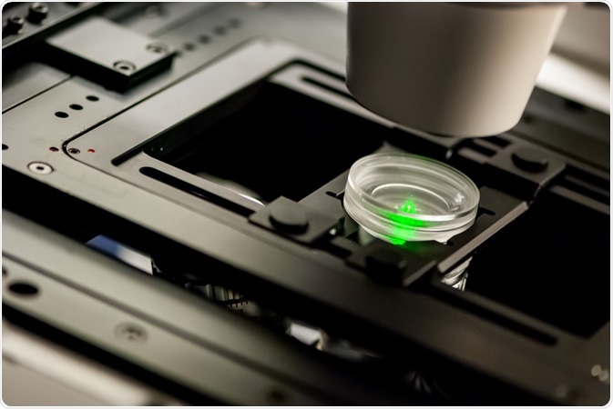
The integration of fluorescent labeling with advanced microscopy techniques has enhanced forensic science investigations and research.
 Image Credit: Micha Weber / Shutterstock.com
How is microscopy used in forensics?
A forensic investigation on determining the cause of death often depends upon the analysis of the evidence of the time since death, otherwise known as the postmortem interval (PMI).
While several macroscopic observations can be utilized to determine the PMI, microscopy techniques, particularly electron microscopy (EM), can provide more accurate PMI estimates. For example, EM has successfully been used to study morphological alterations in human nerves to assess what stage of myelin degradation has occurred.
Several subtypes of EM like transmission electron microscopy (TEM) have also been used to provide information on the cause of death. More specifically, TEM has been used to detect ultrastructural changes in the myocardium and study alterations in skin morphology and mentalization that resulted from suspected cases of electrocution.
In addition to EM, atomic force microscopy (AFM) has also been used for several different forensic investigation applications. Some examples include the characterization of surfaces with potential fingermark deposits, trace evidence analysis of gunshot residues (GSR) and the evaluation of shooting distance.
Applications of fluorescent microscopy
Fluorescent microscopy is associated with several advantageous properties that make it an ideal tool for forensic researchers. Fluorescent microscopy techniques have been widely used within forensic science to assist in the visualization of latent fingerprints, identification of GSR and examination of other types of trace evidence.
Latent fingerprints
Fingerprints are the unique identities of each individual and do not change as an individual gets older, thereby making this type of forensic evidence extremely powerful during criminal investigations. In addition to identifying individuals present at a crime scene, fingerprint analysis is also crucial to preventing and detecting counterfeiting.
Since fingerprints are often invisible to the naked eye, additional efforts by forensic investigators are required to develop and ultimately allow for the visualization of latent fingerprints (LFPs). Several traditional methods that can be used for the development of LFPs include powder dusting, ninhydrin spraying and cyanoacrylate fuming, many of which are simple and effective methods.
Despite their ease of use, many of these methods are associated with high contrast and low background interference, both of which can cause challenges in the accurate visualization of LFPs.
The unique fluorescent properties of several different nanomaterials have led many researchers to incorporate these materials into LFP detection. Quantum dots (QDs), for example, are associated with both exceptional fluorescence intensity capabilities and a large Stokes shift, both of which allow for an enhanced contrast of LFPs when irradiated by ultraviolet (UV) visible light.
Due to the fluorescent label potential of lanthanide phosphors, recent work on this material has led to the fabrication of red-emitting La2(MoO4)3:Eu3+ phosphor microcrystals for the visualization of LFPs deposited onto several different nonporous substrates. Some of the unique characteristics of this novel material include high contrast and low background interference, both of which contribute to the ability of this label to provide well-defined finger ridge details when analyzed by SEM.
Another type of fluorescent label that has been coupled with SEM and TEM include a red and green-emitting CdSiO3:Tb3+/Eu3+ nanopowder, which was fabricated by cetrimonium bromide (CTAB) assisted sonochemical processes. The choice to incorporate rare earth elements like Eu3+ and Tb3+ into this nanopowder was largely due to the capabilities of these ions to emit in the red and green regions, respectively, of the electromagnetic spectrum.
This novel nanopowder is also equipped with silicates, which, due to their stable crystal structure, strong absorption within the near-UV region and impressive long-term durability properties, also enhances its luminescent characteristics.
This novel nanopowder was found to be highly sensitivity and efficient, as well as prevent background hindrance during the microscopic evaluation of fingerprints on various porous and nonporous surfaces ranging from aluminum foil and glass bottles to a teacup and currency note.
GSR
After a firearm has been discharged, the bullet exits the weapon, which causes a mixture of both gaseous and fine particulate matter to be emitted. Taken together, the residue from the primer which ignites the main propellant, as well as any material lost from the bullet and the barrel, can be referred to as GSR or, in more general terms, firearms discharge residue (FDR). Some of the traditional methods for analyzing GSR include the Griess test, which can be used to measure nitrates and sodium rhodizonate for lead; however, these tests are often insufficient in their ability to identify multiple elements with individual particles and are not specific to GSR.
The combined approach of SEM with energy dispersive X-ray spectrometry (SEM-EDX) is a powerful tool that has been used by forensic investigators to detect GSR. Some research has found that incorporating fluorescent lanthanide taggants during ammunition type analyses can assist in SEM-EDX identification. In addition to being used to enhance microscopic evaluation from collected evidence, this type of taggant can also be integrated into monochromated light sources and viewing filters during the initial screening for GSR at the crime scene.
References and Further Reading
Jones, B. J. (2019). Microscopy in Forensic Sciences. In: Springer Handbook of Microscopy. 1507-1524. doi:10.1007/978-3-030-00069-1_32.
Li, F., Liu, S., Qi, R., et al. (2017). Effective visualization of latent fingerprints with red fluorescent La2(MoO4)3:Eu3+ microcrystals. Journal of Alloys and Compounds 727; 919-924. doi:10.1016/j.jallcom.2017.08.182.
Basavaraj, R. B., Nagabhushana, H., Darshan, G. P., et al. (2017). Red and green emitting CTAB assisted CdSiO3:Tb3+/Eu3+ nanpowders as fluorescent labeling agents used in forensic and display applications. Dyes and Pigments 147; 346-377. doi:10.1016/j.dyepig.2017.08.011.
Further Reading
Last Updated: Dec 14, 2020