There are multiple kinds of cell death. Cells can undergo programmed cell death, which is a series of events that undergoes a predictable order at certain times during development.
For example, when your limbs develop, initially the tip of the limb is shaped like a paddle, then the cells in between the digits die and that is what shapes the fingers.
Cell death, known as apoptosis, can also be activated by toxins, or chemotherapeutic agents and other chemicals coming from outside the cell; or it can be activated internally, by the cell assessing its own situation and deciding it’s damaged in some way and that it would be best off just to commit suicide.
For example, cells that detect that their DNA is damaged may activate the apoptotic programme.
There are other types of cell death. For example there is a type of cell death called necrotic cell death. There are many differences between apoptosis and necrosis, although they both result in cell death.
Necrotic cell death involves a different series of morphological changes, which means a different series of events occur in the cell.
A necrotic cell death often results from traumatic injury of the cell, for example if the cell is punctured or there is severe organelle damage.
Overall, there are many types of cell death. The cell deaths can either be beneficial or harmful to the animal.
For example, the nervous system produces a vast excess of cells compared to what it needs, so there is a lot of pruning needed to get the right number of cells.
There are survival factors that are produced in limiting amounts, so only the cells that get enough survival factors go on to live and the others die. This is good for the organism
Another type of cell death that can be beneficial to the animal happens if you are exposed to some carcinogens or radiation and that causes damage to your cells’ DNA. Some cells may then be damaged and some may have mutated, which could lead to the creation of tumors.
It is beneficial for the animal if these cells recognise that they are damaged and they commit suicide rather than going on to become tumor cells. This could be described as a surveillance mechanism which prevents damaged cells from proliferating.
Although cell death can be a good thing, it can also be a bad thing. For example, too much cell death can cause degenerative diseases, such as Alzheimer’s and Parkinson’s disease.
This is because you need a certain number of cells to be alive in order for the body to function properly.
It is important that cell death is tightly regulated so that you don’t get too many or too little cells.
Please could explain to us the process by which toxins normally cause cell death?
Toxins induce cell death by apoptosis. The key event occurs at the mitochondria, which are the energy-producing organelles. The cells have to make a decision on whether to commit cell death or not.
A “toxin” is a very general term, it is not very specific. We use a variety of different toxins that make cells undergo cell death.
One that we commonly use in the lab is ethanol. At very low concentrations ethanol is not toxic, but at high levels it induces cells to undergo apoptosis.
We use other types of chemicals and drugs as well.
The immediate action of each toxin or drug might be different.
For example, ethanol will get metabolized into other products; some of these are oxygen free-radicals.
The toxins ultimately activate a series of enzymes called caspases, which are the master regulators of life and death decisions in the cell.
There are a series of caspases. These are referred to as initiator and executioner caspases. They are enzymes that can cut up other proteins.
Cutting up a protein can either activate a protein or deactivate the protein, depending on the protein. Sometimes they cut off a piece of the protein that is inhibiting it. This is what caspases themselves are like: if you cut them then you actually activate them.
Other proteins are destroyed when you cut them.
With caspases, the initiator caspases cleave and thereby activate the executioner caspases.
The toxins can result in caspase activation, which then sets off a whole series of events in the nucleus, the cytoplasm of the cell, the cytoskeleton, the organelles and so forth.
Essentially, a whole series of events occur downstream of the activation of the caspases. These ultimately lead to cell death.
At what point was it previously thought that cell death due to toxins was irreversible?
The exact point of no return is a bit controversial and it is probably different in different cell types.
There are some known examples of cells that can actually arrest apoptosis after caspase activation. This is different to what we’ve been looking at, however, which is cells that are already far along the apoptosis pathway which then can reverse that series of events. They then repair the damage that has been done.
There are some interesting biological examples of cells that have activated caspases but then don’t die. There is an interesting example, in drosophila (fruit fly) sperm development.
A sperm only really needs its DNA, which it will deliver to the egg; its tail, so that it can swim; and mitochondria so that it can produce energy. It does not need much else.
As drosophila sperm mature they activate the apoptotic pathway, but they do it in a very controlled way. They just get rid of the cytoplasmic constituents that they don’t need anymore, they leave everything else in good shape. Thus they undergo a partial apoptosis.
Aside from a few clear examples like this, in most cells once the caspases are activated, the cells die.
Caspases activation was generally considered a point of no return. But we found that cells could not only activate caspases, but experience the downstream consequences of caspases, yet still recover almost fully.
How did your research originate if it was previously thought that cell death due to toxins was irreversible?
The idea originated with a post-doctoral fellow in my lab, who was a graduate student at the time in Hong Kong. He wondered what would happen if the inducer of cell death was removed.
He was curious as to how far the cells could before they could not recover again.
The idea really came from his experimenting in the lab.
How does your research show that cells can bounce back after toxins have been removed?
One question that people ask is did your research show that just a few cells survived and did not undergo apoptosis? The first thing I say is that it is clearly not the case as we can document that more than 90% of the cells undergo this fairly uniform response once the toxin is removed.
It is not a minority escaper population, but the vast majority of cells in the population that undergo reversal of the events leading to cell death.
The next thing people might ask is whether it is possible for the results to actually be caused by a small minority of cells that grow into a bigger population.
The most compelling experiment that addresses this concern is a time-lapse movie of a single cell which shows the whole cell shrivel up and look like it is going to die; then you can watch it recover once the inducer has been removed. The cytoplasm expands back out; the organelles repair themselves; the nuclei expand again and so forth.
Morphologically you can watch it happen. You can watch one cell or a whole field of 30 cells for example.
We can also show the results at a biochemical level. We have assays that look at whether a cell’s DNA is damaged; whether the caspases have been activated and so forth. We can use this to see how many cells have recovered.
Did the surviving cells have any lasting damage?
There are multiple breaks in the cell’s DNA as they undergo the process of apoptosis. This is because enzymes that cleave DNA are activated.
Once they start to recover they activate DNA repair mechanisms. Most of the breaks in the DNA are sealed and DNA repair fixes most of the problems.
However, in a small percentage of the cells mutations persist. If we do a mitotic chromosome spread, we can see that there are pieces that got joined together that shouldn’t have and so forth.
These obvious chromosomal abnormalities occur in around 1-2% of surviving cells.
There are probably more subtle mutations in a larger amount of cells.
We can see functionally there is an increased amount of oncogenic transformation; that is the ability of the cells to overgrow and the initial signs of transformation into a tumor cell.
Functionally there are mutations that cause these cells to act like cancer cells.
What type of cells did you use in this research and do you think it is applicable for other types of cells?
There are two published papers, one describes the phenomenon in cancer cells, and the more recent paper describes the phenomenon in a number of different types of normal cell.
Most of the experiments in the paper work with a type of fibroblast cell line called anti-age 3T3 cells. These are not cancer cells, but they have a normal karyotype and are considered to be relatively normal fibroblasts. Although they do grow in cultures, so they’re not perfectly normal!
We also saw the results in primary cells, which are cells derived immediately from an animal. For example, primary liver cells are where you take a mouse’s liver and grow its cells. These are clearly normal cells as they are derived directly from animals.
We saw the process in macrophages, which are blood cell types and in cells derived from the heart. The latter is a very interesting observation, as we see a potentially negative consequence of this process as the cells that come out of the recovery may have mutations and might become cancerous.
This process probably evolved in order to be beneficial to an organism. This very strong will to survive may be useful in cells that are difficult to replace.
For example, some of the cells do not self-renew, such as the neurons in the adult brain, the cells in the heart and so forth. These cells are very difficult for an organism to replace.
And so, maybe this very robust survival mechanism exists in order to protect cells and tissues from permanent injury due to a transient exposure to something bad, such as a toxin or chemical. It seems to make sense that it is better for non-renewable cells to be alive, even if they’re a little bit damaged, rather than to be dead.
The research was carried out on cells in mice and rats, do you think it will hold true for human cells?
We have observed the process in one type of human cell: these are a very famous human cervical cancer cell line called HeLa cells.
We haven’t currently observed it in normal human cells, as these are harder to get hold of. However, the observations in HeLa cells along with cells in mice and rats make us optimistic that it will also occur in normal human cells.
In unpublished data we have also seen the process happen in fruit fly cells. Overall, we think that it is probably a very fundamental process and thus is very likely to be conserved in human cells.
What potential medical benefits could arise as a result of this knowledge?
This is the most exciting part as we think the implications will be very wide reaching.
If we can elucidate the mechanism by which cells survive this transient exposure to a toxic agent, then we can imagine two ways that we could derive a medical benefit.
If we developed drugs that enhanced the survival process, we could develop ways to spare neurons in degenerative diseases. This could slow, halt or even arrest the progression of diseases such as Alzheimer’s, Parkinson’s, Huntington’s disease and so forth.
There could also be medical benefits for cancer therapies.
During normal treatment for cancer, we use chemotherapy which induces apoptosis. The problem is we have to give the chemotherapy then take it away, as we don’t want to keep giving it continuously as it is toxic. This means that some of the cells may undergo this process of reversing apoptosis.
If we understood the mechanism of this reversal well enough we may be able to develop additional medication that would prevent the cell survival mechanism during chemotherapy.
We are very excited to pursue this research and elucidate the mechanism which we can then harness to develop new ways for treating degenerative diseases and cancer.
What future plans do you have for research in this field?
There are many things that we would like to do.
First of all, the research we have done so far has all been done outside the animal in cultures; one of our top priorities is therefore to detect the cell death reversal process, which we have named anastasis, in a living organism.
We are turning to the fruit fly for this as we have very advanced genetic tools for this organism.
The problem with this research is that cells that have undergone anastasis look a lot like normal cells! Thus it is hard to tell whether they have been through this process in their history or not.
The live imaging that you can do in the culture dish, where you watch the cells undergo the process, is much harder to do inside of an animal.
The way we are going to look at this involves marking cells so that they permanently express the green fluorescent protein (GFP).
We have done this so far in the fruit fly, we are also developing the same type of tool for the mouse, and we have filed a provisional patent on that approach.
In addition to detecting these events in living animals, it is also important for us to elucidate the mechanism of the process.
We have a good heads start on this as the expression of all the genes in the fruit fly have been examined, so we can look at which go up and which go down during the process. We can also look at what time points these events occur.
We are currently in the process of analysing all this data to determine which pathways are activated and which are inactivated. These are going to be the molecular targets for the medical therapies.
Where can people find more information on this research?
They can find more information in our published paper: http://www.molbiolcell.org/content/early/2012/04/23/mbc.E11-11-0926
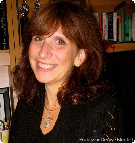
About Professor Denise Montell
Denise is a Professor and Director of the Graduate Program in Biological Chemistry at The Johns Hopkins University School of Medicine.

She started off her career by earning a B.A. degree with honors in Biochemsistry and Cell Biology from the University of California in 1983. Then she completed her Ph.D in Neurosciences at Stanford University in 1988, under the guidance of Dr. Corey Goodman.
She carried out postdoctoral work with Dr. Allan Spradling at the Carnegie Institution of Washington. She moved to an independent “Staff Associate” position at the Carnegie in 1990.
From 1992 she has been a faculty member in the Department of Biological Chemistry at The Johns Hopkins University School of Medicine.