In a just released article in the Journal of Molecular Recognition, Dr. Hermann Schillers et al. report the first visualization of individual microvilli on living cells with atomic force microscopy.
Schillers, H., et al. (2015) PeakForce Tapping resolves individual microvilli on living cells. J. Mol. Recognit., doi: 10.1002/jmr.2510., Courtesy of Bruker Nano Surfaces
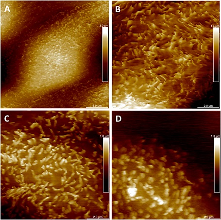
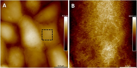
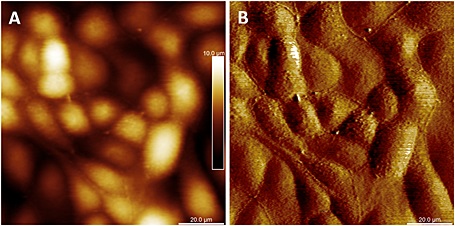
Microvilli are soft, microscopic cellular membrane protrusions present on epithelial cells that act to increase available surface area, enhance transmembrane transport, and serve as mechanosensors. Changes in their density and morphology are associated with diseases, such as celiac disease. As noted by Dr. Schillers, direct observation of microvilli on live cells had long eluded the AFM community.
Dr. Schillers, lead author of the article, noted:
It was previously impossible to resolve the finest structures of a live cell like microvilli, but now with the BioScope Resolve, I can image them easily in one hour. This opens up exciting opportunities for new studies. Observing the structural and therefore functional integrity of microvilli on living cells will help to understand the development of microvilli-dependent diseases.”
Conventional AFMs subject live cells to high normal and lateral forces, yielding images that are dominated by the structure of the harder, underlying cytoskeleton and thus limiting their ability to track any changes in situ on the membrane of live cells.
Schillers, H., et al. (2015) PeakForce Tapping resolves individual microvilli on living cells. J. Mol. Recognit., doi: 10.1002/jmr.2510., Courtesy of Bruker Nano Surfaces
Bruker’s BioScope Resolve™ overcomes this challenge by probing the cell membrane at lower forces and with less spatial averaging from the extended probe structure and its movement through the viscous medium, bringing soft cell membrane structures into clear view for the first time.
Schillers, H., et al. (2015) PeakForce Tapping resolves individual microvilli on living cells. J. Mol. Recognit., doi: 10.1002/jmr.2510., Courtesy of Bruker Nano Surfaces
About BioScope Resolve
BioScope Resolve is an AFM designed specifically for the highest resolution imaging of all biological samples while on the inverted optical microscope. BioScope Resolve is the only AFM to resolve individual microvilli on live cells. It provides the most complete range of capabilities for cell mechanics and molecular force spectroscopy.
Its exclusive PeakForce QNM® and FastForce Volume™ techniques deliver the highest resolution and fastest force mapping capabilities possible with an AFM and the widest range of force distance ramp rates. In addition, BioScope Resolve offers complete synchronization of AFM imaging and force spectroscopy with optical microscopy techniques, enabling new kinds of correlated experiments.
BioScope Resolve is available with a full array of accessories including temperature and environmental control for live-cell imaging.
 Bruker: Launches portable MOBILE-IR II spectrometer