3D imaging technology provides images of the cell cluster down to the nuclei while mitigating drug discovery risks
Olympus Corporation today announced the U.S.-only launch of a new 3D cell analysis technology with ability to accurately analyze 3D cell cultures down to the nuclei, improving the reliability and accuracy of the entire drug discovery process, from compound selection to animal experimentation and clinical trials in humans.
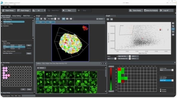
Above: NoviSight™ 3D Cell Analysis Software intuitive user interface provides recognition, analysis, and statistical results in one convenient location
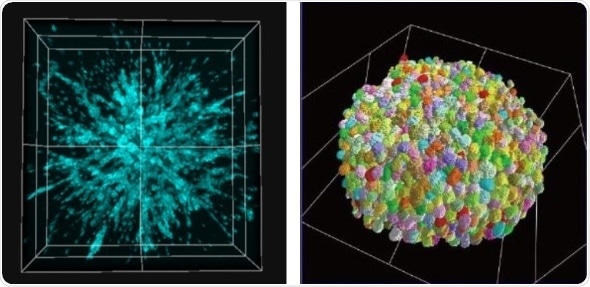
Bottom Left: An example image of cancer remodeling (Invasion Level; High)
Bottom Right: 3D detection result of nuclei in 200 um spheroid
Some photos provided courtesy of Lawrence J.
Ellison Institute for Transformative Medicine of USC
The search for new drugs to treat diseases is a long and costly process with no guarantee of success. Biotechnology and pharmaceutical companies can invest as many as 10 to 20 years and hundreds of millions of dollars in the effort to develop a new drug, yet the probability of success is low; among 30,000 compounds, only one will become a new drug. The use of 3D cell models that simulate a living body help reduce these risks and speed up the drug discovery process. Using 3D cultured cell tissue, known as spheroids and organoids, researchers can analyze the effects and toxicity of new drugs in a bioenvironment similar to that of a living human body. This approach improves the accuracy of results at the preliminary stage of clinical trials.
To develop a solution to meet this need, Olympus combined its 3D imaging technology with powerful algorithms and a new method to analyze the whole cell model in 3D. The result is Olympus’ new NoviSight™ software with True 3D cell analysis technology, a valuable tool designed to help accelerate drug discovery research.
Used with Olympus’ confocal laser scanning microscopes, such as the FLUOVIEW® FV3000 system, NoviSight 3D cell analysis software provides images of the cell cluster down to the nuclei. The software’s True 3D technology uses multiple microplate images to provide accurate morphology data and the ability to quantitatively analyze the effect of medications, including growth suppression and cell survival rates. A range of parameters can be easily and precisely measured, enabling researchers to count the number of cells that have suppressed growth, proliferated, or been annihilated. NoviSight 3D cell analysis software also makes it easy to compare the effects of different medications at various concentrations.
In addition to improved insights and accuracy, NoviSight 3D cell analysis software has practical features built into the user interface that can facilitate and accelerate the interpretation and validation process:
- Recognition, analysis, and statistical results are displayed all on one screen
- Alternate between 3D and 2D views of sample
- Interact with the quantitative data to display it in a scatterplot, heat map, or graph
- Clicking points in one of the graphical display options automatically opens the corresponding original image
- Export data as a CSV or FCS file for further analysis
Launch Background
One of the common methods used in drug discovery is cell analysis. Conventional cell analysis involves using 2D cell culture samples. However, the gaps in structure between 3D cells in the human body and 2D cell cultures result in many drug candidates being determined as incompatible.
The limitations of conventional 2D cell culture analysis affect the reliability and accuracy of the entire drug discovery process, from compound selection to animal experimentation and clinical trials in humans. With the advancement of cell culture technology, working with 3D cell culture samples has become a reality. The ability to accurately analyze 3D cell cultures has the potential to save pharmaceutical manufacturers time and reduce costs. In the U.S., where drug discovery research is especially active, there is a growing demand for 3D cell analysis technology.
_4a0be3d757714179b9f906417b52987b-80x70.jpg) Olympus Discovery Center inaugurated at the University of Maryland
Olympus Discovery Center inaugurated at the University of Maryland