The coronavirus disease 2019 (COVID-19) pandemic, caused by the severe acute respiratory syndrome coronavirus 2 (SARS-CoV-2), has led to intense stress worldwide, threatening human health, life, and the global economy. As such, an unprecedented global research effort is ongoing to find effective measures to contain the virus and bring normalcy back to human life.
Such investigations involve drug development efforts, the identification and isolation of therapeutic neutralizing antibodies specific to the virus, and research into the pathogenesis and management of the condition. An essential condition for such research is the availability of an animal model that recapitulates human infection and disease.
Now, a new preprint research paper posted to the bioRxiv* server reports the development of a modified mouse model that allows productive SARS-CoV-2 infection of the mouse lungs, as well as the exploration of type I and III interferon responses in this condition.
Mice in SARS-CoV-2 research
The SARS-CoV-2 virus binds to the angiotensin-converting enzyme 2 (ACE2) receptor on its target host cell using its receptor-binding domain (RBD) on the viral spike protein. The human ACE2 (hACE2) receptor is thought by many receptors to be the primary entry point for the virus. Though homologous ACE2 receptors are found in mice, they bind poorly to SARS-CoV-2 spike protein, thus preventing productive infection.
Transgenic mice expressing hACE2 under the control of a promoter gene, such as K18 or HFH4, have also been developed. However, it is not simple to use genetically modified (GM) mice for SARS-CoV-2/COVID-19 research.
This has been accomplished using adenovirus vectors (Ad5) or adeno-associated virus (AAV), innocuous viral vectors that can express hACE2 genes within the infected cells of the lungs. The virus has also been adapted for mouse infection, but this infection may not faithfully mirror the effects of therapeutic interventions in humans due to the adaptations.

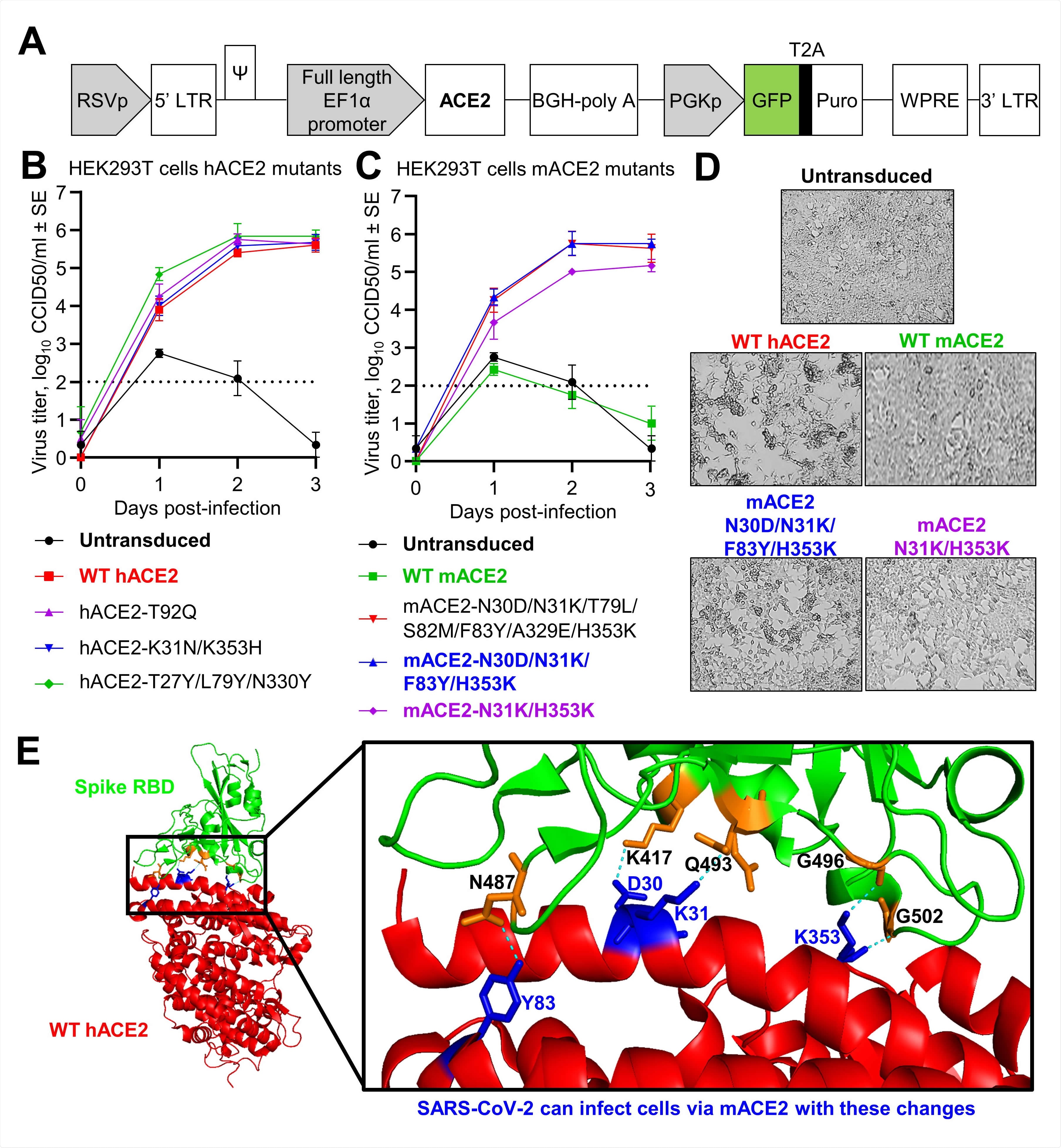
Mutational analyses of human and mouse ACE2 using ACE2-lentiviruses in vitro. A) Schematic of pCDH-EF1α-ACE2-BGH-PGK-GFP-T2A-Puro lentiviral vector. B-C) Growth kinetics of SARS-CoV-2 over a three day time course in HEK293T cells transduced with WT or mutant hACE2 (B) or mACE2 (C) infected at MOI=0.1. Data is the mean of 1 (mACE2-N31K/H353K) or 2 (all others) independent experiments with 3 replicates in each and error bars represent SEM. D) Inverted light microscopy images of HEK293T cells transduced with the indicated ACE2 lentivirus and infected with SARS-CoV-2 at MOI=0.1. Images were taken at 72 hpi and were representative of triplicate wells. E) Crystal structure of the Spike RBD:hACE2 complex (PBD: 6M0J) viewed in PyMOL and highlighting key interactions (blue) between hACE2 (red) and Spike RBD (green).

 This news article was a review of a preliminary scientific report that had not undergone peer-review at the time of publication. Since its initial publication, the scientific report has now been peer reviewed and accepted for publication in a Scientific Journal. Links to the preliminary and peer-reviewed reports are available in the Sources section at the bottom of this article. View Sources
hACE2-lentiviral system
The current study focuses on the use of lentivirus-mediated gene expression in the epithelium of the mouse lung. This system was first used in order to express the cystic fibrosis transmembrane conductance regulator (CFTR) in cystic fibrosis as a therapeutic measure. The success of this method confirmed that it could lead to long-term expression of the therapeutic gene.
The researchers here describe an ACE2-lentiviral system that allows replication-competent SARS-CoV-2 to be cultured in cells in vitro and to infect mouse lungs in vivo. This allows GM mice to be used to study SARS-CoV-2 research, especially to reveal the role of interferons in the induction of inflammation following SARS-CoV-2 infection.
It avoids neuroinvasive infection seen in K18-hACE2 mice. However, the greatest benefit is the incorporation of the lentiviral RNA into the genome of the host cell. This ensures the lentiviral proteins are expressed at a stable level for an extended period.
Studying the binding of the RBD in detail
The scientists found that hACE2-lentiviruses could identify the role of specific residues in the RBD in viral binding. In HEK293T cells, that are naturally non-permissive to SARS-CoV-2 replication as they lack hACE2, transduction with these viruses led to replication.
They tested the role of the N90 glycosylation motif by inserting the T92Q mutation in hACE2. This mutation abolishes this glycosylation site on the receptor, which was predicted to enhance the spike RBD's affinity.
They also introduced the K31/K353 mutations into the receptor, predicted to reduce its affinity for the spike RBD. They found that these mutations did not significantly affect viral replication and thus are apparently not essential for viral infectivity. Instead, the whole of the RBD may be responsible for the tropism exhibited by the virus.
Reduced spike-ACE2 binding may still be enough for productive infection, which is important when designing hACE2 inhibitors.
Mouse ACE2 mutants permissive for SARS-CoV-2 replication
The researchers also discovered that four key residues that take part in the RBD-ACE2 interactions are responsible for the difference in infectivity between cells that carry hACE2 and mouse ACE2, namely, D30, K31, Y83, and K353. Mutants at N31K and H353K allow productive SARS-CoV-2 infection of cells carrying mouse ACE2 receptors. The other two enhance infection to the levels seen with hACE2 infection.
This led to the introduction of the hACE2-lentivirus system into mouse cell lines to establish replication-competent SARS-CoV-2 infection, though the replication level depends on the type of cell.
Mouse models of SARS-CoV-2 infection
The same system was used to explore the role played by type I and type III interferon pathways in SARS-CoV-2 replication. Three GM mouse models that are deficient in these pathways, namely, C57BL/6J, IFNAR-/- and IL-28RA-/- were used.
The transduced C57BL/6J mice showed significant viral titers for all mouse strains, but not the untransduced mouse lungs. Viral titers peaked on day 2 and fell thereafter.
Viral RNA levels were low and fell rapidly in untransduced lungs but were much higher and remained elevated at all time points in transduced lungs. The efficiency of hACE2 expression in transduced lungs is comparable to that achieved by other systems, as with K18-hACE2 mice.
When the lungs of transduced infected C57BL/6J mice and infected controls were compared for innate immune responses. They found distinct interferon and cytokine signatures by day 2 post-infection. This included IL-2, IL-10, IL-6, TNFα, IL-4, IFNγ and CSF3 signatures.
By day 6 post-infection, the inflammation was observed to resolve, with tissue repair well underway as demonstrated by the cytokine profile. This model thus correlates well with the acute inflammation followed by resolution seen with COVID-19.
The Th2 bias and the presence of IL-4 are linked to COVID-19-induced lung damage. Molecular signaling pathways that were upregulated included MAPK1 and MAPK9, while both T and B cells were expressed at higher levels. The anti-inflammatory activity of estrogen appeared to be responsible for the robust upregulation of estrogen receptor 1.
Interferons key to inflammation in COVID-19
Lungs from any of these three models showed comparable viral loads, indicating equivalent levels of viral replication in the presence of deficient type I or III interferon receptor expression. The study showed that type I and III interferon responses are essential to induce the lung inflammation seen in COVID-19, even though interferons do not rise to very high levels following infection.
In fact, the SARS-CoV-2 virus encodes proteins that depress interferon signaling pathways. While type III interferons may be a first-line defense against infection at the epithelial surfaces, type I, which is more powerful, may be triggered by the breach of type III responses. As mentioned already, knockout of type III IFN or type I alone did not stop replication, except in part.
Only if all interferon expression is abolished does viral replication increase. However, knockout of these two pathways led to a lower level of inflammation following SARS-CoV-2 infection.
Vaccine evaluation
This system was also used in wild-type C57BL/6J mice to allow vaccine assessment. Vaccination with either infectious SARS-CoV-2 or its UV-inactivated form led to protective immunity against the infection. This was associated with a marked lymphocytic infiltrate.
Conclusion
The use of lentiviral transduction to introduce hACE2 into mouse lung cells led to the recapitulation of the cytokine profile associated with severe disease, as in other mouse models and following COVID-19 disease in humans.
“We demonstrate broad applicability of this new mouse model in vaccine evaluation, GM mice infection, and in vitro evaluation of ACE2 mutants.”

 This news article was a review of a preliminary scientific report that had not undergone peer-review at the time of publication. Since its initial publication, the scientific report has now been peer reviewed and accepted for publication in a Scientific Journal. Links to the preliminary and peer-reviewed reports are available in the Sources section at the bottom of this article. View Sources