Researchers in the United States have developed a new model for testing mutations in the four structural proteins of severe acute respiratory syndrome coronavirus 2 (SARS-CoV-2) – the agent that causes coronavirus disease 2019 (COVID-19).
Testing of the proteins for their effects on viral assembly, packaging and host cell entry provided a previously unanticipated explanation for the increased fitness and spread of SARS-CoV-2 variants of concern.
The researchers from the Gladstone Institutes in San Francisco in California and the University of California in Berkeley say the platform can be used to test viral variants outside of a biosafety level 3 environment. They add that it is also ideal for developing new SARS-CoV-2 antivirals since it is highly sensitive, quantitative and scalable to high-throughput workflows.
A pre-print version of the research paper is available on the bioRxiv* server, while the article undergoes peer review.

 This news article was a review of a preliminary scientific report that had not undergone peer-review at the time of publication. Since its initial publication, the scientific report has now been peer reviewed and accepted for publication in a Scientific Journal. Links to the preliminary and peer-reviewed reports are available in the Sources section at the bottom of this article. View Sources
Efforts to understand increased fitness have been restricted to biosafety level 3 labs
Understanding the molecular mechanisms behind the increased transmissibility of SARS-CoV-2 variants is essential for the development of therapeutics and vaccines. However, research is hindered by studies being restricted to biosafety level 3 labs.
Efforts to understand the improved fitness of viral variants have so far been limited to studies that use pseudotyped lentivirus particles engineered to display the spike protein. The spike protein mediates the initial stage of the infection process when its receptor-binding domain attaches to the host cell receptor angiotensin-converting enzyme 2 (ACE2).
However, most mutations that occur in circulating variants of concern lie outside of the spike gene and are therefore inaccessible using this approach.
All SARS-CoV-2 variants so far defined by the World Health Organization contain at least one mutation within seven amino acids (N:199-205) of the nucleocapsid protein – a structure that is essential for the binding, packaging, and release of viral RNA.
“Despite its functional importance and emergence as a mutational hotspot, the nucleocapsid protein has not been widely studied because of the absence of simple and safe cell-based assays,” writes Jennifer Doudna and colleagues.
What did the researchers do?
The researchers set out to develop a SARS-CoV-2-based packaging system that would deliver exogenous RNA into cells using virus-like particles (VLPs).
“We reasoned that a process mimicking viral assembly to package and deliver reporter transcripts would simplify the analysis of successful virus production, budding and entry,” says the team.
For such VLPs to successfully deliver reporter transcripts into cells, they need the packaging signal for RNA recognition.

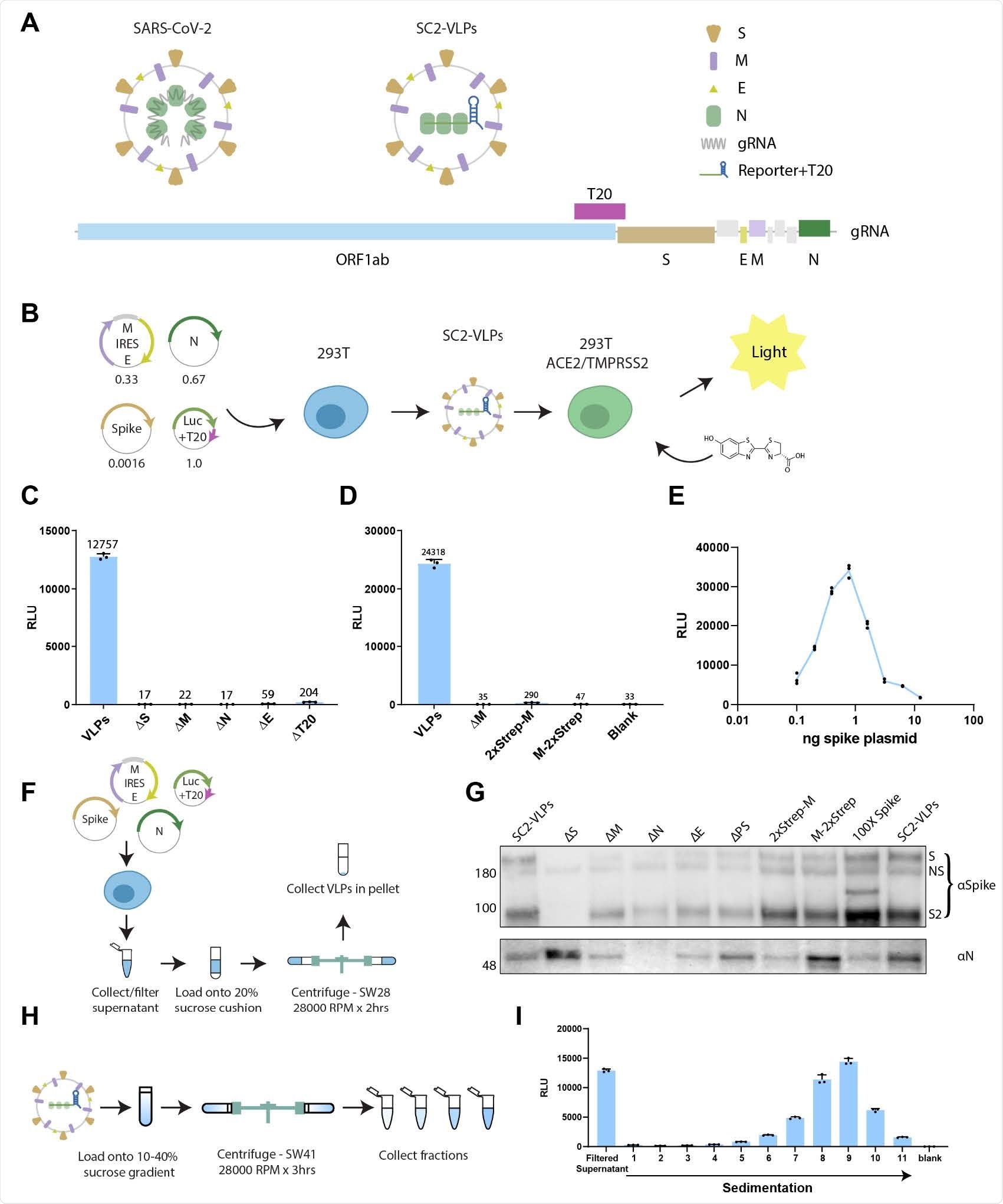
Design and characterization of SC2-VLPs. A) Schematic of SARS-CoV-2 virus and vector design. B) Process flow for generating and detecting luciferase encoding CoV-2 VLPs. Numbers below plasmid maps indicate ratios used for transfection. C) Induced luciferase expression measured in receiver cells (293T overexpressing ACE2 and TMPRSS2) from “Standard” CoV-2 VLPs containing S, M, N, E and luciferase-T20 transcript as well as VLPs lacking one of the components. D) N- or C-terminal streptag on membrane protein abrogates vector induced luciferase expression. E) Optimal luciferase expression requires a narrow range of spike plasmid concentrations corresponding to ~1ng of plasmid in a 24-well. F) Schematic for purification of virus-like particles (VLPs) including CoV-2 VLPs. G) Western blot showing spike and N in pellets purified from standard SC2-VLPs and conditions that did not induce luciferase expression in receiver cells. H) Schematic for sucrose gradient for separating CoV-2 VLPs. I) Induced luciferase expression from sucrose gradient fractions of CoV-2 VLPs.
Based on the reported packaging sequences for SARS-CoV-1 and murine hepatitis virus, Doudna and colleagues hypothesized that the packaging signal for SARS-CoV-2 might lie within the genomic fragment “T20” (nt 20080-22222), which encodes the non-structural protein 15 (nsp15) and nsp16.
They, therefore, designed a transfer plasmid that encodes a luciferase transcript containing T20 and tested for SC2-VLP production by co-transfecting packaging cells (HEK293T) with the transfer plasmid and plasmids that encode the structural proteins.
Supernatant secreted from these packaging cells was incubated with receiver 293T cells that expressed both ACE2 and another host cell entry factor – transmembrane protease serine 2 (TMPRSS2).
The team found that luciferase was only expressed in receiver cells when all four structural proteins (spike, membrane, nucleocapsid and envelope) were present, along with the T20-containing reporter transcript.
Identification of the RNA packaging sequence enabled engineered transcripts to be assembled together with the four structural proteins into VLPs that delivered the transcripts to ACE2- and TMPRSS2-expressing cells.
This provided the opportunity to analyze mutations within all structural proteins and the steps involved in the viral life cycle.
The researchers were surprised by the findings
The researchers were surprised to find that while mutations in spike did not affect particle assembly and cell entry, VLPs bearing any one of the four nucleocapsid mutations that are found universally in more transmissible viral variants exhibited increased particle production and up to 10 times more expression of reporter transcript in receiver cells.
Further analysis indicated that mutations within the linker domain of the nucleocapsid protein improved the assembly of SC2-VLPs, leading to either increased overall production of VLP or a larger fraction of VLPs containing RNA.
“In either case, these results suggest a previously unanticipated explanation for the increased fitness and spread of SARS-CoV-2 variants of concern,” writes the team.
Potential future uses of the new platform
“We present a strategy for rapidly generating and measuring SARS-CoV-2 VLPs that package and deliver exogenous RNA,” says Doudna and colleagues.
The researchers say the strategy is not only useful for understanding the molecular virology of SARS-CoV-2 but also for the development and screening of therapeutics that target viral assembly, budding, maturation and cell entry.
“This strategy is ideally suited for the development of new antivirals targeting SARS-CoV-2 as it is highly sensitive, quantitative and scalable to high throughput workflows,” they write.
The team also says it may be possible to introduce silent mutations within the packaging signal sequence to generate weakened strains of SARS-CoV-2 for use as an infectious vaccine.

 This news article was a review of a preliminary scientific report that had not undergone peer-review at the time of publication. Since its initial publication, the scientific report has now been peer reviewed and accepted for publication in a Scientific Journal. Links to the preliminary and peer-reviewed reports are available in the Sources section at the bottom of this article. View Sources
Journal references:
- Preliminary scientific report.
Doudna J, et al. Rapid assessment of SARS-CoV-2 evolved variants using virus-like particles. bioRxiv, 2021. doi: https://doi.org/10.1101/2021.08.05.455082, https://www.biorxiv.org/content/10.1101/2021.08.05.455082v1
- Peer reviewed and published scientific report.
Syed, Abdullah M., Taha Y. Taha, Takako Tabata, Irene P. Chen, Alison Ciling, Mir M. Khalid, Bharath Sreekumar, et al. 2021. “Rapid Assessment of SARS-CoV-2–Evolved Variants Using Virus-like Particles.” Science 374 (6575): 1626–32. https://doi.org/10.1126/science.abl6184. https://www.science.org/doi/10.1126/science.abl6184.