At the time of publication, I was a post-doc in the lab of Dr. Paola Arlotta in the department of Stem Cell and Regenerative Biology at Harvard University. I did my graduate work at Duke University with Dr. Cagla Eroglu, where I gained my appreciation and fascination with the non-neuronal glial cells of the central nervous system, predominantly astrocytes, and microglia.
As a graduate student, I discovered that astrocyte cells closely associate with neuronal synapses and even regulate how they wire together. I chose to do a post-doc with Paola Arlotta because her lab was at the leading edge of understanding how different neuronal cell types were built in the cerebral cortex. I felt that there were major discoveries to be made by combining my expertise in glial biology with the lab’s expertise in neuronal diversity. Together, we uncovered a code of communication between excitatory neurons and microglia of the cerebral cortex, the region of the brain responsible for higher-order cognitive processes.
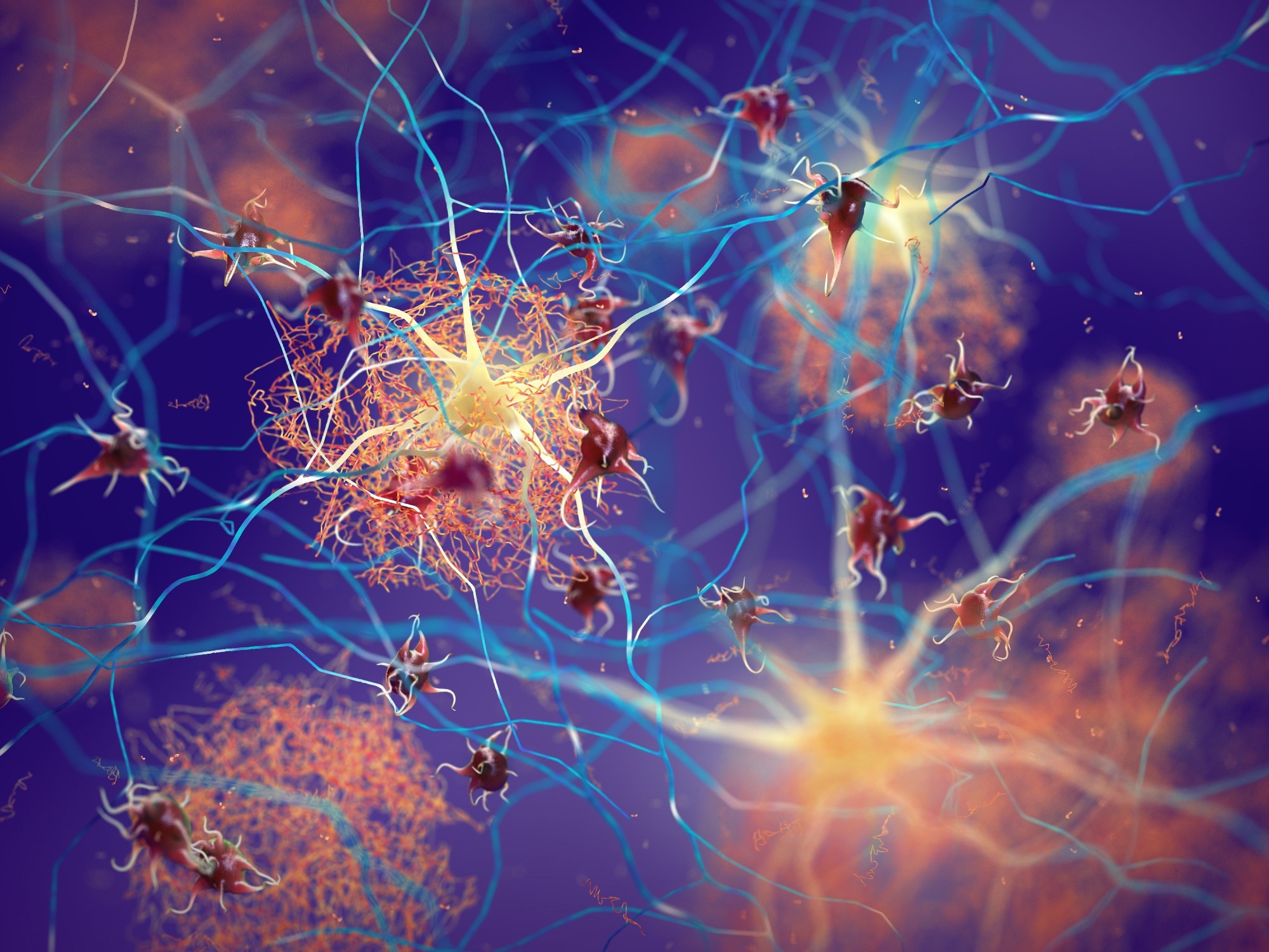
 Image Credit: nobeastsofierce/Shutterstock
Researchers are increasingly learning about the many roles of microglia. What are these tiny immune cells, and how do they play a role in brain function, health, and disease?
Microglia are the local macrophages of the brain, which means they have an immunological cell heritage. In fact, they are derived from a region outside the developing embryo, called the yolk sac, where they migrate through the bloodstream and colonize the brain, eventually remaining there behind the mostly cell-impenetrant wall that is known as the blood-brain barrier.
Historically, microglia have been known to act as cells that eat and remove debris from the brain (i.e. dead cells and clean up after brain damage). However, we are now learning that they do so much more, including sensing and responding to neural activity. They also play outsized roles in human health. Many neurological disorders are linked either directly or indirectly to microglia function, including Autism Spectrum Disorders, Alzheimer’s Disease, and Multiple Sclerosis, just to name a few.
Your latest research suggests that microglia cells can “listen in” to neighboring neurons and change their molecular state to match them. Can you explain what this means and how this occurs?
Microglia, by their immunologic nature, are cells that “listen” and “sense” the environment around them. They possess many small branches that constantly survey their local area to find weak synapses, sites of damage, and evaluate the level of neuronal activity nearby, among other things. We knew from past research that microglia from one brain region express different cell receptors (i.e. the molecules involved in listening) than other brain regions, but it was unclear how this translated at the local level of a single microglia.
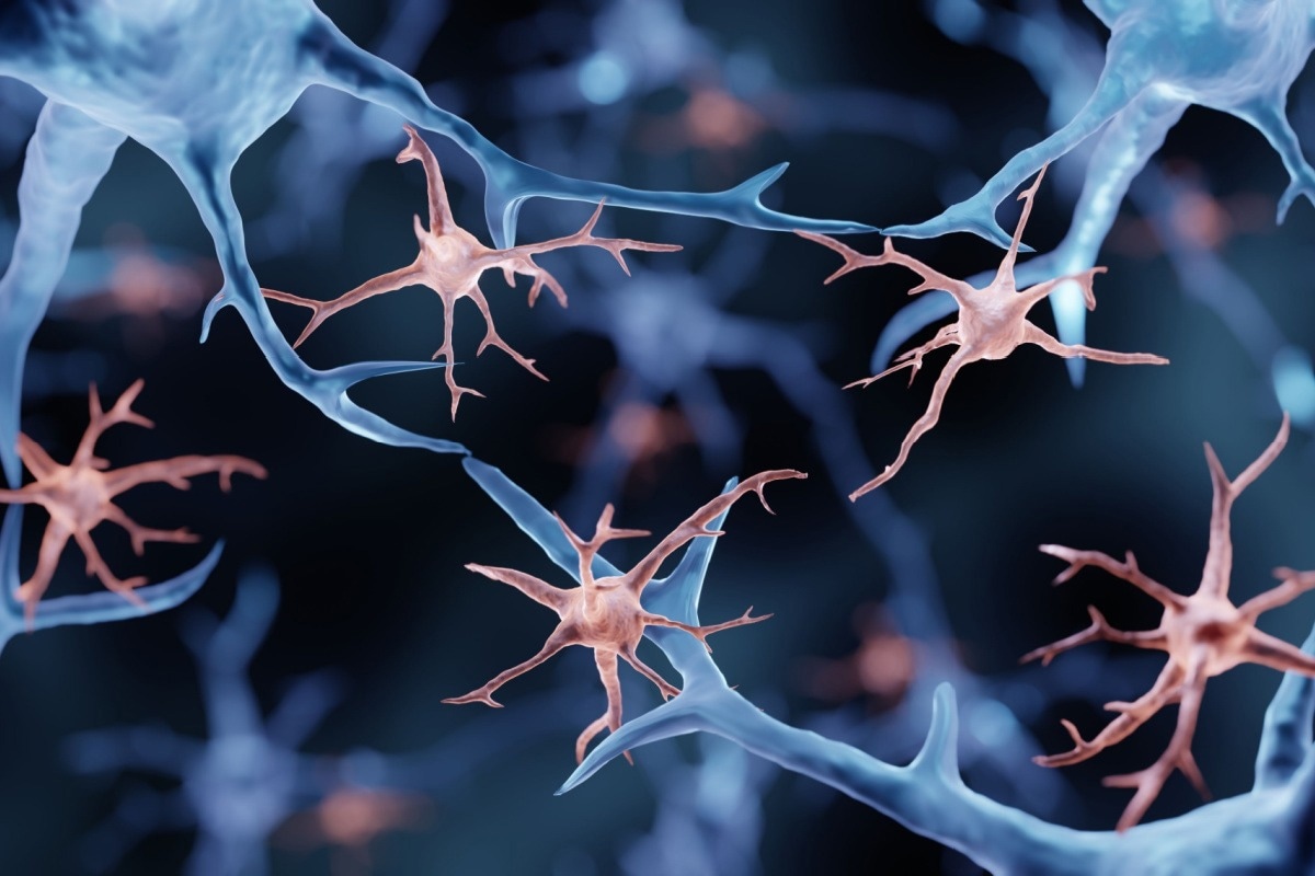
 Image Credit: ART-ur/Shutterstock
We found that within a single brain region (the layers of the cerebral cortex) that houses many different types of excitatory neurons, they can locally control microglia in two important ways: 1) distinct neuron subtypes locally recruit different numbers of microglia to their area, and 2) they “tune” the transcriptional profiles of local microglia, somewhat like a musician tuning an instrument to make the right sound. This latter point is quite important because it suggests that different neurons involved in different brain activities adjust the cellular profile of local microglia to match the needs of their circuits.
We postulate that this is done, in part, by different signaling molecules that are expressed by different classes of excitatory neurons. We found these by profiling the expression of all signaling molecules in neurons and correlating this expression atlas with all of the signaling molecules in different states (or tunes, going back to the music analogy) of microglia. We were stunned to see the amount of specificity in signaling between these major subdivisions of cells.
Your study was conducted by using genetic profiling methods to examine the microglia in the different layers. Can you explain more about how you conducted your research and the findings you uncovered?
We attacked this question in two major ways, by profiling microglia from mice, which is good, although not a perfect correlate to the human brain. In the first approach, we took out the cortex, then carefully micro-dissected the layers of the cortex. We then extracted all of the microglia and profiled them using a power tool called single-cell RNA-sequencing. This method allows researchers to view the RNA expression profile (in other words, the repertoire of expressed genes) of each and every cell in isolation from other cells.
At first, we found that all of the cells from all of the layers that we extracted were microglia by the expression of genes that are unique and specific to microglia. But we then found on top of the base layer of identity existed a secondary layer of gene expression that correlated with the layer from which the microglia were micro-dissected. This gave us the “gene signature” of each layer-enriched microglia state, or the tuned state of the microglia from each layer. It is important to note that each layer of the cortex houses a different subset of excitatory neurons. Thus, we were able to correlate, neuron subtype (by layer) with microglia state (by layer).
The second approach used an even more powerful profiling tool, which allowed us to peer into the transcriptional expression of all cells (neurons, microglia, other glia, etc…) within the intact brain, without having to micro-dissect it. This approach, called Multiplex Error-Robust Fluorescence In Situ Hybridization (or MERFISH for short), was applied to the mouse brain using the gene signatures we found in our first profiling experiment detailed above. By applying this method, we could map, in three dimensions, the exact location of every microglia and every excitatory neuron with exquisite precision.
With this map in hand, we found that microglia states exist in layers as we had found before. More exciting though, we found that each microglia resides within a neighborhood of unique neuron subtypes and that the state of microglia depends on the local composition of its nearby neuron neighbors. This suggests that the level of specificity lies at the level of cellular interactions in neuron-microglia neighborhoods.
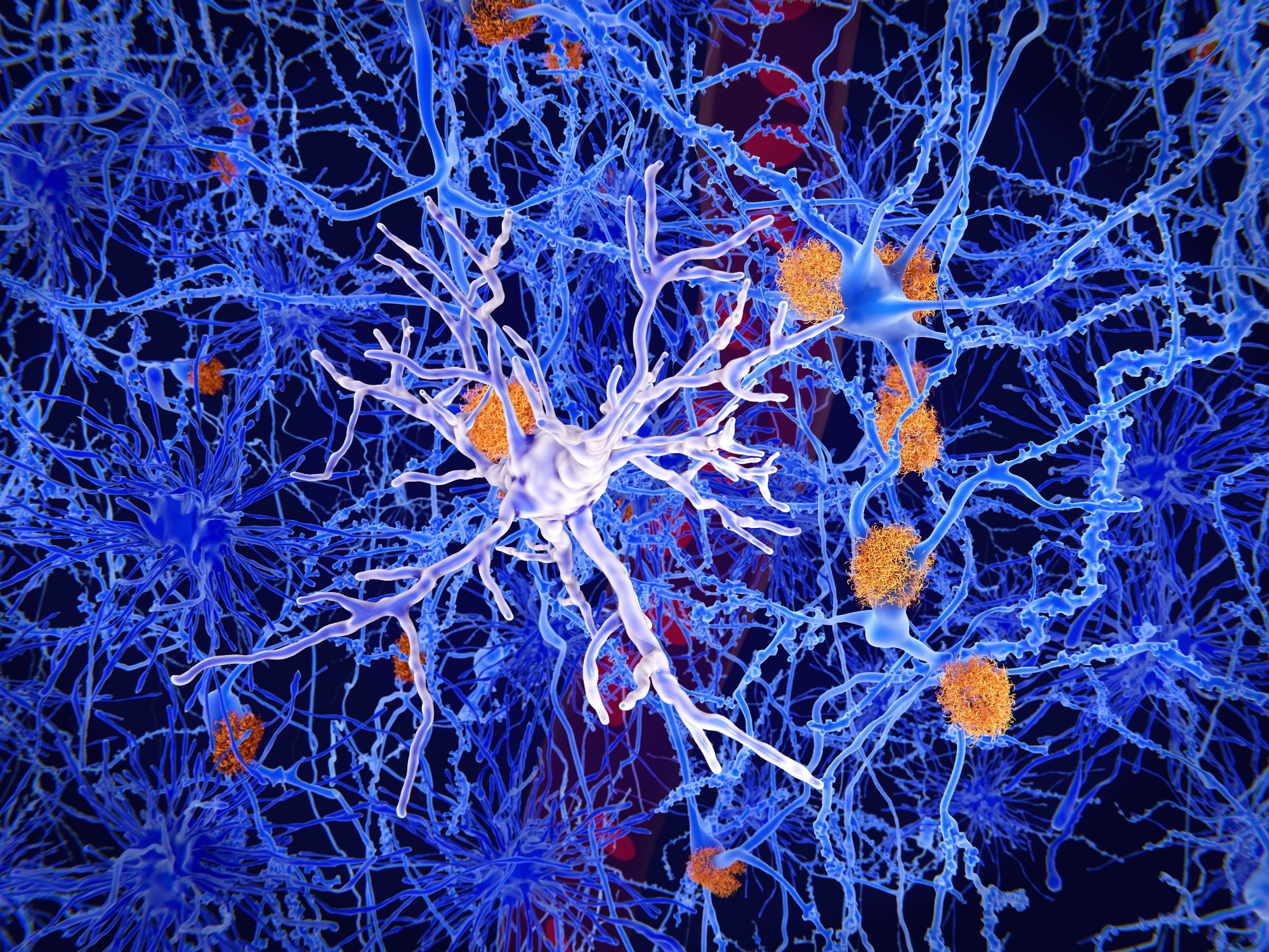
 Image Credit: Juan Gaertner/Shutterstock
What are some of the consequences that occur when the communication between microglia and their neuron partners goes wrong?
Our research did not dive into the effects of miscommunication between microglia and their neuronal partners. However, our signaling atlas presents to the field and wealth of starting points to identify what could go wrong and, maybe more importantly, how we could potentially fix or correct circuits when there is a miscommunication between neuronal subtypes and microglia.
One very interesting note from human research is that there has been identified Autism Spectrum Disorder (ASD) disruptions between upper-layer neurons of the cortex and microglia. Our dataset is primed to be mined to uncover the molecular mechanisms of those upper-layer disruptions in humans with ASD.
How could the results of this new research help open the door for lines of research that can accurately target the communications between microglia and their neuron partners?
Like I mentioned in the previous question, our signaling atlas between neuron subtypes and microglia is a treasure trove of data, waiting to be mined by those in the field of neuroimmunology. Many of the communication signals are pathways that may be “druggable” or can be amended through gene therapy. It’s an exciting time to see how targeting microglia can fix neurons or neural circuits and, ultimately, maybe, neurological disorders.
What are the next steps for you and your research?
I have transitioned to a biotechnology company aimed at using glial cells as a therapy for neurological disorders. I hope the data generated from this published study will be a springboard for other labs to identify how to produce different microglia states in culture for testing, analyses, and therapies. I also hope it can help derive new insights into mechanisms of neurological disease initiation or progression.
Where can readers find more information?
Readers can find the original study here:
About Jeffrey Stogsdill, Ph.D.
Currently, I am a senior scientist at Sana Biotechnology, trying to find ways to use glial cells as a therapy for neurological disorders. For the research conducted on the paper in discussion, I acted as a post-doc in the lab of Paola Arlotta in the Department of Stem Cell and Regenerative Biology at Harvard University. Extensive bioinformatic analyses were performed by Kwanho Kim in the lab of Joshua Leven at the Broad Institute of Harvard and MIT. The project was carried out with funds from the NIH and Broad Institute of MIT and Harvard (through Paola Arlotta and Joshua Levin) and funds from HHMI (Jeff Stogsdill).
