Laboratory rats were subjected to third-degree burns over 30% of their total body surface area (TBSA) and divided into sham, case, and control groups. The experimental methodology comprised immunofluorescence (IF) staining, immunohistochemistry (IH), and TUNEL staining for burn and atrophy visualization, and Western blotting, qRT-PCR, and metagenomic sequencing for gut microbiome characterization and evaluation.
Study findings revealed that burn injury results in persistent and severe muscle wasting, weight loss, and protein breakdown. These observations can be traced to the gut microbiota, specifically alterations to the typical ratio between abundances of Firmicutes and Bacteroidetes (F/B ratio). Inulin was found to support and increase Firmicutes- and reduce Parabacteroides distasonis abundances, stabilizing the gut microbiome and reversing burn-induced weight loss and skeletal muscle atrophy.
 Study: Dietary supplementation with inulin improves burn-induced skeletal muscle atrophy by regulating gut microbiota disorders. Image Credit: Dorota Milej / Shutterstock
Can the gut microbiome treat burns?
Burns, especially severe third-degree ones, are known to cause substantial and persistent skeletal muscle degeneration and, more recently, have been found to have profound and long-term effects on gut microbial assemblies. Skeletal muscle wasting is one of burn pathophysiology's central factors contributing to both recovery time and post-recovery symptom severity. This wasting is aggravated by stress, systemic inflammatory response, and reduced physical activity levels, resulting in delayed burn healing.
The gut microbiome, also called 'gut flora' and 'gut microbiota,' refers to the complete assemblage of bacteria, bacteria, archaea, fungi, and viruses that inhabit the digestive tracts of animals. The gut microbiome plays a critical role in health, with alternations in community composition resulting in substantial effects, especially in the neuronal, digestive, and cardiovascular systems. Recent research has uncovered the role of burns in altering diversity and abundance changes in gut microbiota, resulting in a notable decrease in probiotic genera and a corresponding increase in opportunistic pathogens.
Previous animal model experiments have depicted that burns can contribute to microbial community dysbiosis and intestinal barrier dysfunction. Unfortunately, no studies have yet explored the link between microbial dysbiosis and burn-induced muscle atrophy, leaving the mechanistic underpinning of this interaction unknown.
Inulins are a clade of naturally occurring sugars (polysaccharides) belonging to the class fructans (fruit sugars). Previous research has reported the modulatory effects of these compounds in regulating Bacteroidetes and Bifidobacteria microbiome populations. Separate work has elucidated Inulin's potential anti-inflammatory and muscle-loss-attenuating properties, highlighting it as a probable safe and natural intervention to improve burn victims' quality of life (QoL).
About the study
The present study aims to investigate the impacts of inulin supplementation on gut microbiota dysbiosis and muscle atrophy in burned rats. Male Sprague‒Dawley rats (mean age = seven weeks; mean weight = 195 g). Rats were divided into four experimental groups, namely 1. Sham burn (S), 2. Burn (B), 3. Intervention (I), and 4. Validation (V). All groups (except S) were subjected to anesthesia, following which 30% of total body surface area (TBSA) third-degree scald wounds were administered.
Four days following the experiment, rats were euthanized to allow for histochemical evaluations. These evaluations included immunofluorescence (IF) staining to detect myofiber borders in the tibalis anterior (TA), extensor digitorum longus (EDL), and gastrocnemius (GAS) muscles. Immunohistochemistry (IH) investigations were then carried out to estimate the relative expression levels of ZO-1, occludin, and claudin proteins from excised tissue samples.
The TUNEL (terminal deoxynucleotidyl transferase dUTP nick end labeling) staining assay was used to detect the DNA breaks formed when DNA fragmentation occurs in the last phase of apoptosis. Apoptic cell characteristics were recorded and analyzed using high-resolution photography and ImageJ, respectively.
Protein and metagenomic sequencing analyses were employed to characterize real-time gut microbial composition and metabolite secretions and monitor any changes over the study period. These included quantitative real-time polymerase chain reaction (qRT‒PCR), Western blot, and the enzyme-linked immunosorbent assay (ELISA).
Study findings
Examinations of daily rat weight, food intake, muscle weight, and fat weight revealed that groups B and V presented significant body weight loss for the first six days following burn procedures, following which weight was observed to rise gradually. Weight loss was highest in the B group, representing 7.28% of baseline weight. In contrast, Inulin was shown to have a protective effect against weight loss, with the I group only experiencing a 2.94% weight reduction during the same period. Following sham (no) burning, the S group presented a slow upward trend of weight gain.
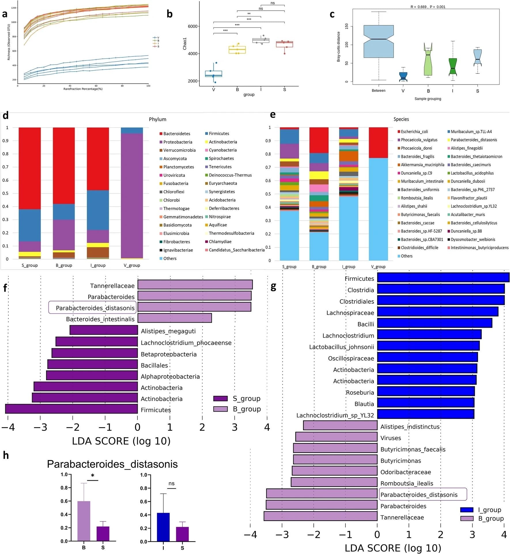 Taxonomic analysis of the gut microbiota in different treatment groups. (a) Rarefaction curve analysis in four groups. (b) Alpha diversity of the four cohorts at the species level, measured in terms of the Chao1 index. (c) Beta diversity of the four cohorts at the species level. (d) Bar plot of taxonomy analysis between four groups in taxonomic levels of phylum. (e) Bar plot of taxonomy analysis between four groups in taxonomic levels of species. The horizontal axis is the group, and the vertical axis is the relative abundance of certain species. (f, g) LEfSe analysis LDA value information at each taxonomic level between different groups. The ordinate shows species with significant differences in LDA values greater than the present value (2.0) in different groups, that is, biomarkers with significant differences. The length of the bar in the chart represents the impact of significantly different species. (h) P. distasonis species relative abundance in different groups. The P value was determined by a two-tailed Wilcoxon rank-sum test, and data are presented as the means ± SEMs; * p < 0.05. S sham, B burn, I intervention, V validation, LEfSe linear discriminant analysis effect size, LDA linear regression analysis, P. distasonis Parabacteroides_distasonis, SEM standard error of the mean.
Food intake evaluations reported increasing consumption trends across groups B, I, and V. Inulin's benefits were further apparent, with the highest food consumption noted in the I cohort, suggesting that the sugars may indirectly hasten burn recovery. Gene- and protein-level assays found an almost threefold higher expression of Muscle atrophy-related F-box (MAFbx) and Muscle atrophy-related F-box (MuRF1) in the burn groups compared to the sham cohort. However, the I group was found to have significantly lower MuRF1 and MAFbx levels than their other burn cohorts, suggesting that Inulin attenuates protein expression in the ubiquitin-proteasome signaling pathway.
Cytokine and intestinal tight junction (TJ) expression levels were further altered by inulin supplementation. Severe burns were found to result in substantially lower intestinal TJ, occludin, and ZO-1 expression levels, but these values were significantly dampened and comparable to S H-scores in the I group. Rarefaction analysis elucidated this phenomenon by highlighting that Inulin increases the alpha diversity of gut microbial communities and helps those undergoing dysbiosis to return to baseline.
"Compared to the burn group, Inulin increased the relative abundance of Firmicutes and Actinobacteria (30% and 3.3%, respectively) in the intervention group and decreased the abundance of Proteobacteria (9.7%). We observed significant differences in the relative abundance of species across treatment groups. These results indicate the differences in the gut microbial communities of rats in different treatment groups."
Functional analysis of gut microbial communities compared against the Kyoto Encyclopedia of Genes and Genomes (KEGG) database presented that Inulin was able to alter the functional annotation of gut microbiota, resulting in higher expressions of metabolic pathways involved in amino acid biosynthesis. Inulin further promoted skeletal muscle protein synthesis through altered phosphatidylinositol 3-kinase/protein kinase B (PI3K/AKT) signaling.
Conclusions
The present study investigates the association between gut microbial communities and skeletal muscle atrophy following severe burns. Study findings indicate that burns have a powerful disrupting effect on gut microbial assemblages, resulting in altered systemic-level protein expressions. Inulin, a plant-derived polysaccharide, was shown to reverse these adverse impacts, promoting burn healing and attenuating skeletal muscle atrophy.
"These insights represent a critical first step and highlight the need to assess whether interventions targeting the gut microbiota could be a strategy for treating burned skeletal muscle atrophy. Given the limited efficacy of current therapies and the considerable impact of skeletal muscle atrophy on the prognosis and quality of life of burn patients, innovative strategies to alleviate skeletal muscle atrophy are urgently needed."
Journal reference:
- Gao, Shan, et al. "Dietary Supplementation with Inulin Improves Burn-induced Skeletal Muscle Atrophy by Regulating Gut Microbiota Disorders." Scientific Reports, vol. 14, no. 1, 2024, pp. 1-14, DOI: 10.1038/s41598-024-52066-8 https://www.nature.com/articles/s41598-024-52066-8
 Scientists uncover shared gene signatures that reveal how mammals age