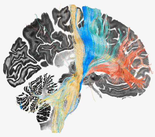
Mass General Brigham researchers identified sets of connections that are disrupted and malfunctioning as a consequence of Parkinson’s disease, dystonia, obsessive compulsive disorder and Tourette's syndrome.
 Fiber bundles associated with symptom improvement following deep brain stimulation in Parkinson’s disease (green), dystonia (yellow), Tourette’s syndrome (blue),and obsessive-compulsive disorder (red). Image Credit: Barbara Hollunder
A new study led by investigators from Mass General Brigham demonstrated the use of deep brain stimulation (DBS) to map a ‘human dysfunctome’ — a collection of dysfunctional brain circuits associated with different disorders. The team identified optimal networks to target in the frontal cortex that could be used for treating Parkinson’s disease, dystonia, obsessive compulsive disorder (OCD) and Tourette's syndrome. Their results are published in Nature Neuroscience.
“We were able to use brain stimulation to precisely identify and target circuits for the optimal treatment of four different disorders,” said co-corresponding author Andreas Horn, MD, PhD, of the Center for Brain Circuit Therapeutics in the Department of Neurology at Brigham and Women’s Hospital and the Center for Neurotechnology and Neurorecovery at Massachusetts General Hospital. “In simplified terms, when brain circuits become dysfunctional, they may act as brakes for the specific brain functions that the circuit usually carries out. Applying DBS may release the brake and may in part restore functionality.”
Connections between the frontal cortex in the forebrain and basal ganglia, structures located deeper in the brain, are known to control cognitive and motor functions. If brain disorders occur, these circuits may become affected, and their communication may become overactive or malfunction. Previous studies have shown that electrically stimulating the subthalamic nucleus, a small region in the basal ganglia that receives inputs from the entire frontal cortex, can help alleviate symptoms of these disorders.
To understand this relationship better, the authors analyzed data from 534 DBS electrodes in 261 patients from across the globe. Of this cohort, 70 patients were diagnosed with dystonia, 127 with Parkinson’s disease, 50 with OCD and 14 with Tourette’s syndrome. Using software developed by Horn’s team, the researchers mapped the precise location of each electrode and registered results to a common reference atlas to compare locations across patients. The researchers used computer simulations to map tracts that were activated in patients with optimal or suboptimal outcomes.
Using these results, they were able to identify specific brain circuits that had become dysfunctional in each of the four disorders, such as those mapping to sensorimotor cortices in dystonia, the primary motor cortex in Tourette’s, the supplementary motor cortex in Parkinson’s disease and parts of the cingulate cortex in OCD. Notably, the identified circuits partially overlapped, implying that interconnected pathways are disrupted in these disorders.
Further, the investigators were able to apply these findings to fine tune DBS treatments and demonstrate preliminary improved results in three cases, including one at Massachusetts General Hospital, a founding member of Mass General Brigham. This patient, a female in her early 20s, was diagnosed with severe, treatment-resistant OCD involving obsessions about food and water intake, along with compulsive skin picking. Following electrode implantation and targeted stimulation, the researchers were able to show a significant improvement in her symptoms one month after treatment.
Except for the three patients that were tested prospectively, the study was a retrospective analysis of data aggregated from multiple centers. Further studies are needed to validate findings in prospective fashion.
We can take this technique further and finely segregate dysfunctional circuits in order to have greater impact with treatment. For example, with OCD, we can look at isolating circuits for obsessions versus compulsions and so on.”
Barbara Hollunder, MSc, Lead Author, Movement Disorders and Neuromodulation Unit, Department of Neurology, Charité – University Medicine Berlin
Source:
Journal reference:
Hollunder, B., et al. (2024) Mapping Dysfunctional Circuits in the Frontal Cortex Using Deep Brain Stimulation. Nature Neuroscience. doi.org/10.1038/s41593-024-01570-1.