LumitTM Immunoassays are a quick and easy alternative to traditional immunoassay procedures such as sandwich ELISAs and Western blots. LumitTM assays are sensitive, have a wide dynamic range, and may be performed in as little as 30 minutes, making them an appealing alternative to time-consuming ELISA and Western blot methods.
Advantages of Lumit™ assays over conventional immunoassays
- Straightforward add-mix-read protocol with no washing steps
- Sensitive luminescence sensing with a broad dynamic range
- Measurement of analytes directly on the cell culture plate or on medium taken from the cells
- No immobilization of plates, beads or other surfaces required
How Lumit™ immunoassays work
NanoLuc® Binary Technology (NanoBiT®) underpins LumitTM Immunoassays. Antibodies are chemically labeled in LumitTM Immunoassays with the small and large subunits of NanoLuc® Luciferase, designated as SmBiT and LgBiT, respectively.
When the two antibodies come into contact with an analyte, SmBiT and LgBiT create an active enzyme and produce a brilliant luminescence signal.
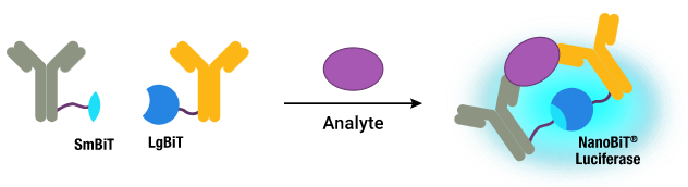
Image Credit: Promega Corporation
Video Credit: Promega Corporation
What are Lumit™ immunoassays?
Lumit™ immunoassays are available as ready to use kits or tools to create your own Lumit™ immunoassay.
The LumitTM Immunoassay Labeling Kit is employed to label antibody pairs for LumitTM Assays. There are also LumitTM Detection Reagents and pre-labeled LgBiT and SmBiT secondary antibody conjugates available.
LumitTM Immunoassays for cytokine detection use a straightforward, no-wash assay technique to quantify target analytes in cell culture samples. The assays can directly run on cell culture samples or on media transferred from cell plates.
The LgBiT and SmBiT subunits are linked to a pair of secondary antibodies from two distinct species in the LumitTM Immunoassay Cellular System. Seeded cells are lysed in a multiwell plate using a NanoBiT®-compatible lysis buffer (digitonin), and the target protein is identified by adding an antibody mix that includes two primary antibodies against the protein as well as SmBiT- and LgBiT-conjugated secondary antibodies.
The LumitTM FcRn Binding Immunoassay is a competitive assay used to assess the interaction between human FcRn and Fc proteins, such as antibodies. The tracer is a Human IgG1 tagged with LgBiT (Tracer-LgBiT). The target is a Streptavidin-SmBiT-bound C-terminal biotinylated human FcRn.
Tracer-LgBiT binds to the hFcRn-SmBiT target in the absence of an antibody analyte sample, producing the strongest luminescence signal. Unlabeled IgG will attempt to bind to the FcRn target in competition with Tracer-LgBiT, causing a concentration-dependent reduction in the luminescent signal.
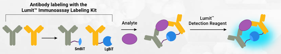
Image Credit: Promega Corporation