This article and associated images are based on a poster originally authored by Johannes Rudolph, Petra Stojanovich and Carl Peters and presented at ELRIG Drug Discovery 2026 in affiliation with University of Colorado Boulder and BMG LABTECH.
This poster is being hosted on this website in its raw form, without modifications. It has not undergone peer review but has been reviewed to meet AZoNetwork's editorial quality standards. The information contained is for informational purposes only and should not be considered validated by independent peer assessment.

PARP1 participates in the initial response to DNA damage and is a treatment target for some cancers. HPF1 was recently identified as a PARP1 cofactor, requiring re-evaluation of existing PARP1 inhibitors; inhibitor association was measured using a kinetic fluorescence polarization assay on the CLARIOstarPlus.
Introduction
PARP1 (poly-[ADP-ribose] polymerase 1) acts in the early stages of DNA damage response. It recognizes and binds single- or double-strand DNA breaks, then becomes active, using NAD+ to add PAR (poly-[ADP-ribose]) chains to itself, DNA, and nuclear proteins like histones.
A function of autoPARylation is the release of PARP1 from DNA. The cancer therapies that target PARP1 inhibit NAD+ association and thus maintain the PARP1 DNA interaction, promoting cytotoxicity.
Recently, a PARP1 co-factor HPF1 (histone PARylation factor1) was discovered that changes the substrate specificity of PARP1, leading to preferential histone PARylation. This newly discovered interaction has spurred the re-evaluation of existing PARP inhibitors (PARPi), which were previously characterized for their association with PARP1 alone.
Here we describe an assay that can address either PARP1 or PARP1-HPF1 complex associations with PARPi using a kinetic fluorescence polarization readout and reagent injections performed by the CLARIOstarPlus.
Assay principle
The kinetic fluorescence polarization assay employed here (Figure 1) exploits the fact that all current PARPi share the same binding site as NAD+.
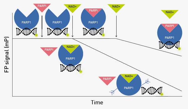
Fig. 1. PARPi association assay. Image Credit: Stojanovic et al.
A fluorescein-labeled double-stranded 18-mer* serves as the tracer. When the 18-mer* is associated with PARP1 (or the PARP1-HPF1 complex), the FP signal is high. PARPi is added for different times before the addition of NAD+, which induces autoPARylation of PARP1 and dissociation of the 18-mer*, observed as a decrease in FP.
If the incubation time for the PARPi is sufficient for it to associate with PARP1 or the PARP1-HPF1 complex, then the FP signal will remain high for longer due to NAD+ access to the binding site being blocked by PARPi.
Materials and methods
- Black 384-well plates (#3575, Corning)
- p18-mer*(5’-phosphate-fluorescein-GGGTTGCGGCCGCTTGGG-3’) (double-stranded; IDT)
- Olaparib (#S1060, Selleck)
- CLARIOstarPlus (BMG LABTECH)
- Other chemicals and reagents were obtained from commercial sources
Experimental procedure
PARP1 and HPF1 were prepared as previously described (50 mM Tris-HCl (pH 8.0), 50 mM NaCl, 1 mM MgCl2, 0.1 mM EDTA). 0.01% IGEPAL was used as binding buffer.
PARP1 (20 nM), p18-mer* (10 nM), and HPF1 (900 nM), when used, were incubated in binding buffer for 15 minutes. Olaparib (20 nM) and NAD+ (6 mM) were dissolved in binding buffer and used to prime reagent injectors 1 and 2, respectively. PARP1 complexes from above were transferred to a microplate (10 μl/well)
Instrument settings
Source: BMG LABTECH GmbH
| . |
. |
. |
| Optic settings |
Fluorescence Polarization, well mode |
| Filters |
Ex: 482–16
Em: 530–40 |
| Gain |
1540 |
| Focal height |
5.9 |
| General settings |
Flashes |
40 |
| Kinetic window 1 settings |
Number of intervals |
15 |
| Interval time |
1 s |
| Injection 1 |
Volume |
10 μl |
| Pause after injection |
1.5–150 seconds |
| Injection 2 |
Volume |
10 μl |
| Kinetic window 2 settings |
Number of intervals |
80 |
| Interval time |
One second |
Results and discussion
As an example of the data collected, the association of the PARPi Olaparib is presented. Figure 2 shows the representative traces of FP signal over time comparing the effect of different delay times before NAD+ addition on the rate of FP decrease associated with the association of Olaparib with PARP1.
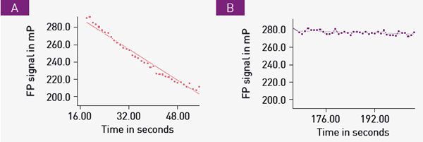
Fig. 2. Olaparib association with PARP1. Image Credit: Image courtesy of Johannes Rudolph et al., in partnership with ELRIG (UK) Ltd.
Following treatment with Olaparib, NAD+ was added after a delay of 1.5 seconds (A) or 150 seconds (B), respectively. An average of two progress curves is displayed along with a linear fit line.
The slope of the linear fit indicates the rate of p18mer* release. As expected, longer delay times before NAD+ addition exhibit a slight decrease in FP signal due to association of Olaparib with PARP1.
As a consequence, less free PARP1 is available to utilize NAD+ and subsequently release p18mer*. Additional experiments were conducted with different delay times to obtain additional rate values. Figure 3 shows a plot of the various rate values versus the delay time before NAD+ addition.
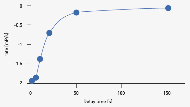
Fig. 3. Replot of linear rates versus delay time. Image Credit: Image courtesy of Johannes Rudolph et al., in partnership with ELRIG (UK) Ltd.
Results presented elsewhere show the linear replot in Figure 3 can be used to discover the apparent rate of association (kobs) for Olaparib. Similar experiments were conducted at different Olaparib concentrations and kobs plotted versus Olaparib concentration.
These results revealed a linear dependence of kobs on Olaparib concentration for PARP1. The same tests were performed using the PARP1-HPF1 complex, which also revealed a linear relationship between Olaparib concentration and kobs. This linear relationship is indicative of a simple one-step association mechanism.
Furthermore, from this plot, the true association rate (kon) for Olaparib can be determined.
Other PARPi were tested with the same association tests. Importantly, these results showed that the same simple one-step association mechanism is employed for all tested PARPi when associating with either PARP1 or the PARP1-HPF1 complex. Figure 4 shows the comparison of the different inhibitors and their association (kon) with PARP1 or PARP1-HPF1 complex.
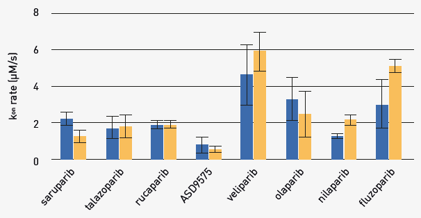
Fig. 4. Comparison of kon for PARPi with PARP1 or PARP1-HPF complex. Image Credit: Image courtesy of Johannes Rudolph et al., in partnership with ELRIG (UK) Ltd.
Average kon (+/-SD) for indicated PARPi with PARP1 (blue bars, left side) or PARP1-HPF1 (orange bars, right side) [n=3-4]. Differences after HPF1 addition are seen for fluzoparib, niraparib, and saruparib.
Conclusion
The kinetic FP assay employed here will be a useful tool in fully characterizing the mechanism of action of PARPi. This understanding should help to further improve inhibitor performance and possibly reveal novel therapies that improve cancer treatments.
About BMG LABECH
 BMG LABTECH has been committed to producing microplate readers for more than twenty years. By focusing on the needs of the scientific community, the company’s innovative microplate readers have earned the company the reputation of being a technology leader in the field.
BMG LABTECH has developed a wide range of dedicated and multi-mode microplate readers for life sciences applications and high-throughput screening.
About ELRIG (UK) Ltd.
The European Laboratory Research & Innovation Group (ELRIG) is a leading European not-for-profit organization that exists to provide outstanding scientific content to the life science community. The foundation of the organization is based on the use and application of automation, robotics, and instrumentation in life science laboratories, but over time, we have evolved to respond to the needs of biopharma by developing scientific programmes that focus on cutting-edge research areas that have the potential to revolutionize drug discovery.
Comprised of a global community of over 12,000 life science professionals, participating in our events, whether it be at one of our scientific conferences or one of our networking meetings, will enable any of our community to exchange information, within disciplines and across academic and biopharmaceutical organizations, on an open access basis, as all our events are free-of-charge to attend!
Our values
Our values are to ensure the highest quality of content and make it readily accessible to all. We will also strive to be an inclusive organization, serving a diverse scientific network. In addition, ELRIG will always be a volunteer-led organization, run by and for the life sciences community, on a not-for-profit basis.
Our purpose
ELRIG is a company whose purpose is to bring the life science and drug discovery communities together to learn, share, connect, innovate, and collaborate on an open-access basis. We achieve this through the provision of world-class conferences, networking events, webinars, and digital content.
Sponsored Content Policy: News-Medical.net publishes articles and related content that may be derived from sources where we have existing commercial relationships, provided such content adds value to the core editorial ethos of News-Medical.net, which is to educate and inform site visitors interested in medical research, science, medical devices and treatments.
Last Updated: Oct 12, 2026
 VANTAstar Microplate Reader from BMG LABTECH