Neuroanatomy is the study of the relationship between structure and function in the nervous system. Neuroanatomy includes the study of macroscopic and microscopic structures. Macroscopic structures are larger structures, such as folds of the brain. On the other hand, microscopic structures include those at the cellular and molecular level, like interactions between neurons and glia.
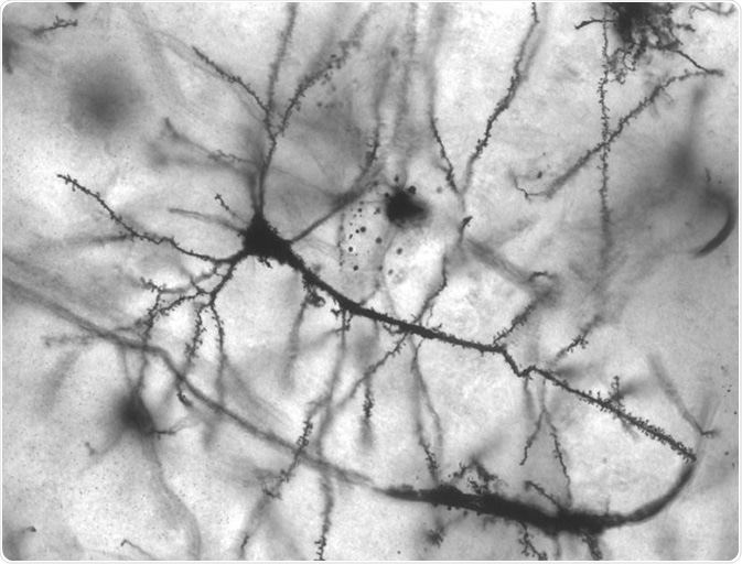
Image: Golgi stained pyramidal neuron in the hippocampus of an epileptic patient. 40 times magnification. Credit: Methoxyroxy~commonswiki/ commons.wikimedia.org.
History of the field
The Edwin Smith Surgical Papyrus, from ancient Egypt, represents the first known record of a neuroanatomy study. It dates to around 1600 BCE. It was the Greek philosopher Alcmaeon who first understood that it is not the heart, but the brain in charge of human body and the senses. Alcmaeon’s work dates to the 5th century BCE. Other scientists and philosophers have built on those works. For example, in the 17th century Thomas Willis (who was a professor and neuroanatomist at Oxford University) published Cerebri Anatome, which is still considered the foundation text of neuroanatomy.
The nervous system
Three components form the foundation of the nervous system: neurons (or nerve cells), neuroglia (glial cells) and extracellular constituents. Neurons process information by sensing the environment, communicating through neurotransmitters, and originating our thoughts and memories.
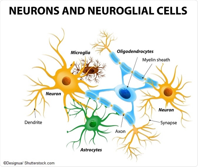
Neurons have a cell body (soma) and two types of extensions, or processes. One is called a dendrite, and the other one is an axon. Neurons usually have more than one dendrite and one axon. Dendrites receive signals which they then send towards the cell body. The axon also transmit signals, but over longer distances.
Neurons form synapses with each other, which can be seen under a microscope. These are junctions where signals are passed from the axon of one neuron, to a dendrite or cell body of another neuron.
There are many types of glial cells. They perform a variety of functions such as maintaining homeostasis, producing myelin, and protecting neurons. Glial cells are interspersed between nerve cells. The main types of glial cell are astroglia, oligodendroglia and microglia.
The nervous system is comprised of fiber bundles that originate from the brain, but also from the spinal cord. These fibers are called nerves, and they are made of neurons plus membranes that separate them into fascicles. Moreover, they form branches to reach every part of the body.
In vertebrates, the nervous system is commonly divided into central part (or central system) and peripheral part (or peripheral system). The central nervous system (often abbreviated as CNS) entails the brain, the spinal cord and the retina, and spinal cord, whereas the peripheral nervous system (often abbreviated as PNS) contains all the other nerves that connect the CNS to the remainder of the body.
Humans have 31 pairs of spinal nerves emanating from the spinal cord. Twelve pairs of nerves emanate from the brain. One of those, the vagus nerve, descends into the trunk and innervates the inner organs (along with other fibers that come from the spinal cord).
The PNS, in turn, is comprised of somatic (controls muscle movement) and autonomic (controls involuntary actions) nervous systems. The somatic system contains neurons which are afferent and which gather sensory data from the environment in order to deliver it in the central nervous system. It also contains neurons which are efferent and that convey motor commands to muscles.
On the other hand, the autonomic nervous system is made of sympathetic and parasympathetic systems. These two systems regulate the body’s basic functions like heartbeat and breathing. Autonomic nerves also habors both afferent and efferent neuron fibers.
Neuroanatomy terminology
A number of standard terms are in use to indicate orientation of structures in neuroanatomy, in reference to the axis of the body or brain. Some of these are:
- dorsal: the top side (from Latin dorsum, back)
- ventral: the bottom side (from Latin venter, belly)
- rostral: the front part of the body (from Latin rostrum, nose)
- caudal: the tail end or back part of the body (from Latin cauda, tail)
- medial: close to the median plane of the body (from Latin medius, middle)
- lateral: away from the median plane of the body (from Latin latus, side)
A separate set of terms is used for orientation planes or sections:
- sagittal: a division relating to the sagittal suture joining the right with the left parietal bones found in the skull
- transverse: a section through any extended structure that is orthogonal towards the axis of the structure, as in a ring-shaped segment of a finger
- coronal: coronal planes or sections parallel with the face, relating to the coronal suture of the skull
- horizontal: being aligned with the horizon
- axial: aligned with the axis of the body
Further Reading
Last Updated: Jun 14, 2019