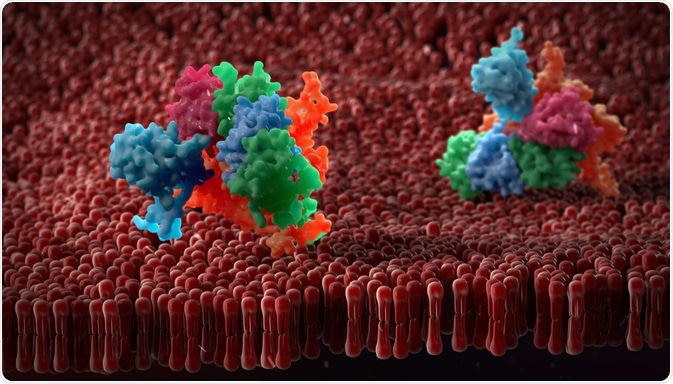
All proteins are made of basic secondary structure units, either α-helix or β-sheets, determined by hydrogen bonding between the amino acids within a peptide chain. On a larger scale, structures are formed by the combination of these secondary structures, and these can form supersecondary structures known as “motifs”.
 urfin | Shutterstock
Motifs have been used as a guide for protein structure for many years. At first, it seemed that proteins which have a similar function shared a resemblance in their 3D shapes, the notable example being hemoglobin and myoglobin. However, in 1973, it was shown that proteins with different functions can also be similar in structure, when Rao and Rossmann identified the nucleotide-binding domains in flavodoxin and lactate dehydrogenase.
What are the basic motifs of proteins?
The simplest motifs are composed of multiple secondary structure units. These simple motifs can contain α-helices and β-sheets that are layered adjacent to one another either in the same direction (parallel) or in the opposite direction (anti-parallel). The simplest motif is the formation of a “loop”, known as a β-turn if it is short, while an unstructured connection is termed a “coiled region”.
Triple helix motif
This is where three α-helices are further twisted into a single coil, or helix. This motif is associated with collagen but has been found in proteins associated with the immune system and host defense.
Four helix bundle
This is where four α-helices are packed together, commonly in a lengthwise direction. This motif can be associated with other motifs to form the overall protein structure or be isolated from other motifs. This motif has been observed in a variety of proteins, including human growth hormone, apolipoprotein and interleukins.
Another example is the combination of β-sheets.
Hairpin
This is a very simple structure, consisting of two antiparallel β-sheets joined by a loop. There can be more than one hairpin connection to form a larger motif.
Greek key motif
This motif also consists of several anti-parallel β-sheets, however, one of the connections between these β-sheets is not a hairpin connection.
Parallel β-helix
This is where parallel β-sheets fold together to form a right-handed coil or helix. Proteins containing this motif include pectate lyase and the tail spike of the phage virus, P22.
β-roll
This motif is similar to the parallel β-helix, however, it is distinguished by being a di-β-strand motif – the parallel β-helix is a tri-β-strand motif. An example of a protein with a β-roll is alkaline phosphatase.
Are there more complex motifs?
These α-helices and β-sheets are not only joined by loops and β-turns but can also be joined by a β-sheet or an α-helix. Metal ions can also play a role in the formation of motifs.
TIM barrel
TIM (triose phosphate isomerase) barrels are where parallel β-sheets are joined by α-helices. This is a widespread motif and was first identified in triose phosphate isomerase, hence the name!
Zinc finger motif
Zinc finger motifs are a group of motifs rather than a single motif. All of these motifs utilize a zinc ion to stabilize the motif structure. In the classical zinc finger motif, two antiparallel β-sheets and an α-helix are brought together by the zinc ion.
These “ββα” units form finger-like projections, which gave rise to their name. Classic zinc fingers are associated with proteins which bind to DNA. Other categories of zinc fingers include nuclear hormone receptors and GAL-4 related proteins.
MegaMotifBase
The motifs listed above are only a subset of known motifs. As implied above, motifs are an important part of proteins, and having information available about these motifs would be of benefit to those attempting to engineer a protein or those wishing to find the function of a new protein.
Pugalenthi and co. developed a database, MegaMotifBase, which contains a database of information on important protein motifs.
Further Reading
Last Updated: Apr 5, 2019