There are several different types of electron microscopes, including the transmission electron microscope (TEM), scanning electron microscope (SEM), and reflection electron microscope (REM.) Each of these types of the electron microscope will be described in more detail in this article, including the benefits and disadvantages of each.
Transmission electron microscope (TEM)
The transmission electron microscope is the original type of electron microscope, which directs a high voltage electron beam towards the specimen to illuminate it and create a magnified image of the sample.
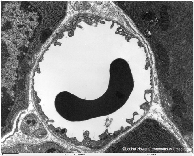
The image shows the cross section of a capillary with a red blood cell present, taken using a transmission electron microscpe.
An electron gun is used to produce the electron beam. The gun is usually fitted with a tungsten filament cathode, which is the source of the electron beam. An anode is used to accelerate the electron beam, and electrostatic and electromagnetic lenses help to focus the beam.
When the electron beam passes through the specimen, it is scattered and provides an image of the microscopic structure of the specimen, which can be viewed through the objective lens of the microscope.
The spatial variation can be examined by projecting the image onto a fluorescent zinc sulfide coated screen. Another method that can be used to record the image is to place a photographic film into the electron beam, which will record the image. A digital camera can also be used to display the image on a computer screen in real time.
Spherical aberration has traditionally limited the resolution of transmission electron microscopes. However, recent developments have helped to overcome this issue to increase resolution with hardware correction of the spherical aberration. As a result, it is now possible to produce images with a resolution below 0.5 angstrom and more than 50 million times magnifications.
The most significant limitation of the transmission microscope is the requirement for very thin specimen samples, usually less than 100 nm. As a result, most biological specimens need to be chemically fixed and dehydrated, in order to be embedded in a polymer resin so that it can be viewed with a TEM.
Scanning electron microscope (SEM)
The scanning electron microscope used a technique known as raster scanning to produce magnified images of the specimen. It directs a focused electron beam across the rectangular area of the specimen, which loses energy as it passes through. The energy is converted into other forms of energy, such as heat, light, secondary electrons, and backscattered electrons. This information can be translated to view the topography and composition of the original specimen.
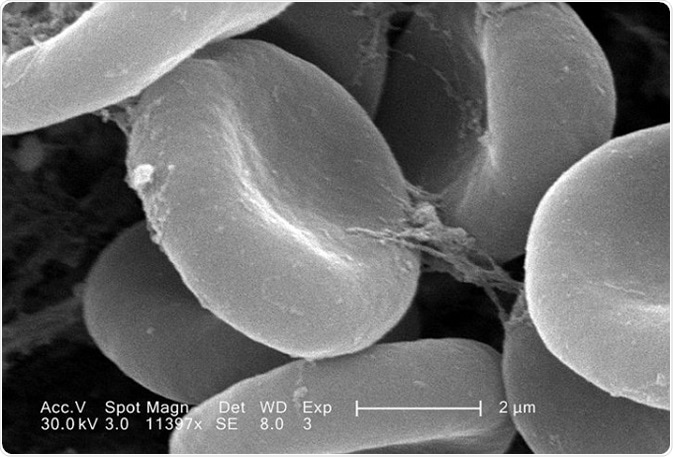
The image shows red blood cells, taken using a scanning electron microscpe.
The resolution of scanning electron microscopes tends to be poorer than that achieved with a transmission electron microscope.
However, it is beneficial because it utilizes surface processes and can hence create images of large samples, up to several centimeters in size, and has a greater depth of field. As a result, the images from an SEM can be good representations of the real shape of the specimen.
Additionally, a specific type of SEM known as environmental scanning electron microscope (ESEM) is able to image samples that are wet or contained in gas. This increases the range of possibilities for which the microscope can be used.
Reflection electron microscope (REM)
The reflection electron microscope involves the detection of a beam of elastically scattered electrons that is reflected off of the specimen that is being examined. The reflection high-energy electron diffraction (RHEED) and reflection high-energy loss spectroscopy (RHELS) techniques are often used in this type of microscopy.
References
- https://www.fei.com/introduction-to-electron-microscopy/types/
- http://www.newworldencyclopedia.org/entry/Electron_microscope
- http://www.keyence.com/ss/products/microscope/bz-x700/study/principle/002/index.jsp
- https://www.sciencelearn.org.nz/resources/502-types-of-electron-microscope
Further Reading
Last Updated: Aug 23, 2018