An interview with Dr. Frank Lafont, Institut Pasteur de Lille conducted by April Cashin-Garbutt, MA (Cantab)
Please can you give a brief introduction to your research?
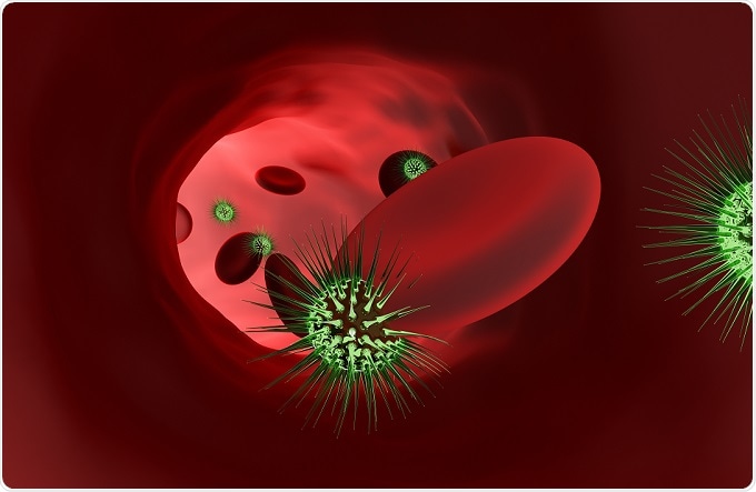
My group is interested in host pathogen interaction and we mainly focus on how bacteria enter cells. In essence, the idea is that if you can prevent bacteria from entering cells, then you will prevent illness.
 Credit: Kotin/Shutterstock.com
How has AFM directly advanced or helped your research?
When a bacterium binds to a cell, which is the first step (the adhesion step), there are a lot of mechanical interactions within that, which then influence how the cell will respond to the invasion by the bug. We think that this is a crucial step that organizes the fate of this response and whether a pathogen is successful at replicating within the cell.
Previously, AFM was used mainly by physicists but now, AFM is increasingly used by MDs and biologists.
Very importantly, when you are an MD or a biologist and you perform, for example, PCR or any other kind of experiment, you want to generate the same results as your colleague in another lab. That was not the case in the AFM field.
People were perfectly aware of all the caveats of the instruments and they would discuss all the parameters that could be changed and the different ways of conducting experiments and they understood each other perfectly.
However, in medicine and biology, you need values that can be trusted and that can all be produced by anyone, anywhere in the world, irrespective of the machine that is used. That was missing and this procedure is used to really calibrate everything so that people have more or less the same values after doing their experiments, irrespective of the system used, the brand of the instrument and whoever is using the instrument.
This is crucial, especially for MDs, if they want to say to someone "you have cancer because you have this amount of values of whatever." That value must be very well-defined, which is something that was missing in the field.
I think it's really a big step forward for MDs and biologists to use AFM, which was not the case before, as it was not a priority instrument in the toolbox of an MD or a biologist.
Increasing Repeatability for Bio-AFM with SNAP
Increasing Repeatability for Bio-AFM with SNAP from AZoNetwork on Vimeo.
How does the standardized nanomechanical AFM procedure (SNAP) eliminate previous sources of error?
The SNAP procedure is based on the calibration of the system, especially the cantilever, which is the crucial part in the AFM instrument. This is where our main interest is focused − on having a perfectly calibrated instrument we can use to do experiments.
This was verified on amorphous gels and on living cells and it was very interesting having these two kinds of samples to experiment on and this was done such that everything was prepared in a wet lab and then dispersed across all the labs in Europe.
People were doing their experiments on these two kinds of samples, on their own instruments, with their own people and at the end, we were finding more or less the same values, which was good.
What is the biggest impact that AFM has made to the biological and nanomedicine research fields?
I think that so far, the main research impact has been in oncology research. Cancer cells are softer than healthy cells, which is true for AFM and for other techniques people are using.
Recently, a review summarized all the results and irrespective of the technique used, it was a common trait that cancer cells were softer. This is maybe the best or most well-known example so far.
How has Bruker technology helped or advanced AFM in biological research?
Bruker was one of the first manufacturers to make AFM available to the biologist. Initially, it was not a perfect machine, but a lot of improvements were made and now they have a system that can be used by biologists or MDs alike.
The first machine was designed such that it was placed onto an inverted microscope and for MDs and biologists, especially cell biologists, it is vital to see cells, and for that, you need a microscope.
What is the importance of meetings, like the AFM Biomed Conference, to you and the AFM research community?
It is really important to gather people who perform different experiments and use different systems and instruments. This is also really the point at which people would decide, for instance, whether to go for a particular EU grant or use the kind of methodology that we were discussing before, the SNAP procedure. This was really made possible thanks to this kind of AFM Biomed Conference.
What direction do you see, or would like to see, AFM going in the next five years? What do you see as the next big thing for AFM?
I remember that I gave a conference maybe four or five years ago and I was asked what the future of AFM would be - I think my answer at that time is still valid today.
There were two main paths. The first one was high-speed AFM. For me, that is useful for what I call “machine at works,” which means looking at a molecular complex and observing the kinetics of that complex. It may be on support of bilayer on cells, but it's really reduced to these kind of small systems, owing to the limitations of high speed AFM, small field and so on.
The second path was correlative microscopy, which combines an AFM with another instrument. It could be a fluorescence microscope and for this to have the same resolution, it would need to be a super resolution system. All these technologies were acknowledged by the Nobel Prize, but it could also be Raman imaging, electron microscopy or any other imaging technique that can not only be combined with an AFM, but combined such that there is a special correlation. The same resolution can be achieved now with fluorescence microscopy, which has reached nanometer resolution.
Where can readers find more information?
About Dr. Frank Lafont

Frank Lafont had medical, scientific and management training and graduated from the Paris VI Pierre & Marie Curie University and from the ESCP-Europe Business School. Regarding Life Sciences, he did his Biology Master practical in Pr J Glowinski’s Laboratory (Neuropharmacology) at the Collège de France (Paris, France), and his Life Science Thesis at the École Normale Supérieure (Paris, France) in Pr A Prochiantz ‘ Laboratory (Developmental Neurobiology). He did his post-doc training at EMBL (Heidelberg, Germany) in Pr K Simons’ Group (Cell biology).
Then, he moved to Geneva (Switzerland) as Junior Lecturer at the University Medical Center in Pr FG van der Goot’s Laboratory (Microbiology) before becoming Scientific Collaborator at EPFL (Lausanne, Switzerland) in S Catsicas ‘Group (Brain and Mind Institute). While at EPFL, he developed his expertise in Atomic Force Microscopy at the Complex Mater Physics Dept (G. Dietler‘s Group).
Since September 2005, he is Group Leader at the Lille Pasteur Institute (Cellular Microbiology and Physics of Infection Group, www.cmpi.cnrs.fr) and since 2010 he is the Scientific Director of the BioImaging Center Lille (bicel.org). In 2014, he was nominated Scientific Officer (Chargé de mission) for microbiology and infectious diseases at the Executive Office of the Biological Science National Institute – CNRS.
Frank Lafont is coordinating a number of Scientific Programs funded by the French National Agency incl. within the Investment for the Future Program and involved in several EU Programs. He is the Vice-President of the French Club for autophagy, Secretary of the French Society for Cell Biology and member of the Scientific Board of the French Society for Biophysics. He is member of the ASCB, Biophys. Soc. and the ASM. He is lecturer and/or responsible of teaching units in Infectious diseases at the Medicine Faculty, and in Biology and Physics Masters at the Science Faculty of the Lille Univ.
His main research interest is to unveil how biophysical properties of membrane can regulate autophagy cell signaling responses induced upon host-pathogen interaction. He has 68 articles in peer-reviewed journals in PubMed and more that 4700 citations.