Following radiation therapy to the head for brain tumors, more than 80% of patients who live six months at least after the treatment report a loss of some cognitive functions such as memory, thinking and related tasks. Now a new study published in the journal Scientific Reports shows why this happens, which in turn could pave the way to correct this damage and protect the brain in future patients.
The researchers found that when exposed to radiation, the brain’s immune cells tend to become like overenthusiastic amateur gardeners entrusted with pruning shears for the first time. They then cut off, not just unnecessarily profuse nerve connections, but even those that are crucial in providing normal access between different brain regions.
The immune system in the brain is constantly engaged in pruning the highly intricate network that links the millions of nerve cells in the brain. This is a normal part of, and even a necessary part of, healthy brain activity. It helps to set up newer and more efficient pathways in place of older and weaker links, as the individual learns and experiences more things in the course of time. This is called neuroplasticity and is mediated by both synaptic and structural plasticity.
However, says researcher Kerry O’Banion, “When exposed to radiation, these cells become overactive and destroy the nodes on nerve cells that allow them to form connections with their neighbors.”
Microglia
The microglial cells that act as the immune sentinels of the brain normally patrol this organ for any signs of infection, injury and tissue damage. They not only detect but respond to these events in a timely fashion, signaling other immune cells and physically removing the debris from the injured site to promote proper healing and minimal scarring. When activated, microglia express complement receptor 3 at high levels and can then shed C3 proteins. This triggers the complement cascade which in turn brings about inflammation and immune reactions. C3 is the meeting point of all the complement activation pathways and its activation is necessary to control multiple white cell functions required for immunity.

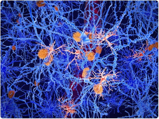
Microglia cells (red) play an important role in the pathogenesis of Alzheimer's disease. Microglia are specialised macrophages that restrain the accumulation of amyloid (orange plaques). 3d rendering. Image Credit: Juan Gaertner / Shutterstock
However, microglia go beyond this traditional role to play an integral part in the constant process of refining and upgrading the connections between neurons, as the brain develops, matures and grows ever more complex. This ongoing editing is a crucial part of memory, cognition, learning and sensory function. C3 is localized on neural synapses and is important in enabling microglial pruning and phagocytosis of unwanted matter during brain development. C3-dependent phagocytosis can slow down the pace of learning and hinder memory development in young and old mice.
Microglia connect closely with neurons at the synapses, those specialized structures in the nerve cell where the axon or dendrite of one nerve cells connect with that of another. Dendrites are the arm-like extensions that reach out from the neuron cell body to receive the axons of other neurons and are therefore rich in synapses. The axon is the long nerve cell extension that transmits messages to another neuron downstream of the first, at the synapses. Obviously, axons from multiple neurons can connect with the same dendrite at different synapses.
When a synapse is in active use, it is maintained in good condition. However, when the synapse falls out of use, a signaling protein is produced that labels that synapse as unnecessary and calls upon the microglia to destroy its link with the other neuron.
The study
The current study focused on nerve networks in the hippocampus because this brain region is closely related to learning and memory.
The current study was done in mice exposed to radiation doses similar to those that are given during radiation therapy. Previous studies in rodents showed that with such exposure, the brain microglia became active and removed dendritic spines, important synaptic structures that mediate neuronal connections by acting as one side of the junction. Without these spines, the nerve cell cannot form a new connection with another neuron.
Interestingly, the researchers also found that new spines were more likely to be removed, which could be a possible hindrance to the formation of new memories. This could be why many patients experience cognitive difficulties following cranial radiation therapy.
Male mice seemed to be more severely affected than female mice after radiation, with respect to cognitive function. The sex of an animal is known to be a prime factor in determining how it develops and how it responds to aging, injury and disease. Microglial activation, synaptic changes and cognitive deficits are different in males and females.
In the current study, microglial activation was more marked in male mice, but the morphology appeared to remain unchanged.
Implications
The results of the study show that even as advanced protocols are being developed to allow more precise radiation therapy and limit collateral damage to healthy parts of the brain, the risk of actual brain damage remains a significant factor in these patients.
Moreover, it indicates two possible ways to meet this challenge. One, nerve cell damage could possibly be prevented using molecules that inhibit the CR3 receptor, which is involved in the removal of synapses by microglia. When such an approach was used in mice, synaptic loss was not observed even with radiation exposure. The second possibility is that the patient may be put on immunosuppressant drugs during the period of cranial irradiation to keep the microglial hyperactivation from occurring.
Journal reference:
Hinkle, J.J., Olschowka, J.A., Love, T.M. et al. Cranial irradiation mediated spine loss is sex-specific and complement receptor-3 dependent in male mice. Sci Rep 9, 18899 (2019) doi:10.1038/s41598-019-55366-6, https://www.nature.com/articles/s41598-019-55366-6