From gut-primed T cells to microglial signaling and persistent gene-regulatory states, researchers map an intricate immune network that connects the brain with the rest of the body.
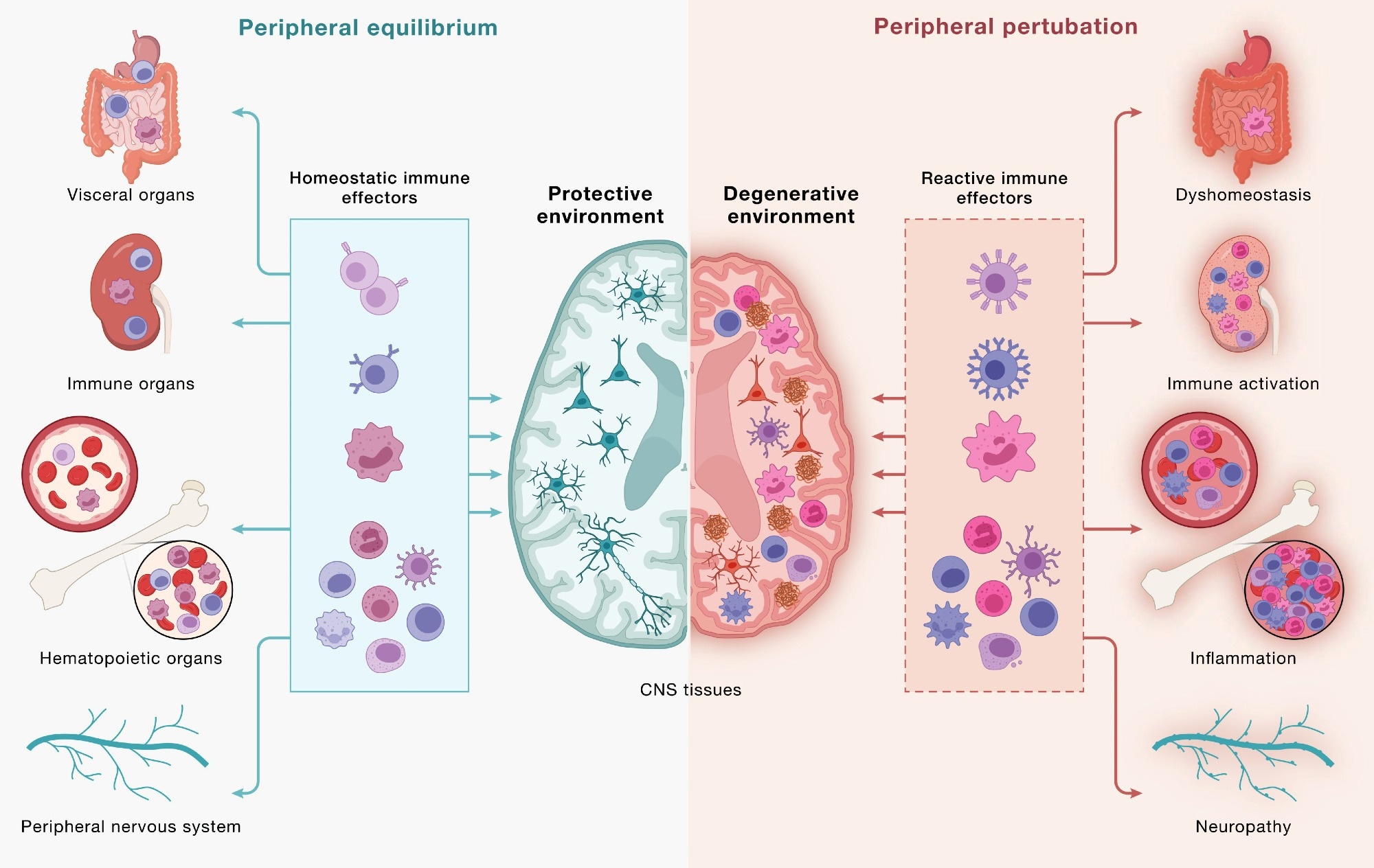
Immune cells communicate across a continuous, bidirectional circuit between the CNS and periphery. Immune cells from across the body engage CNS tissues and, in turn, are shaped by signals returning from the brain. (Left) Under peripheral equilibrium, immune effectors mobilized from visceral organs, secondary lymphoid organs, hematopoietic tissues, and the peripheral nervous system (PNS) - including CD4+ and CD8+ T cells, B cells, monocytes (top 3 cell types), NK cells, dendritic cells, mast cells, neutrophils, and innate lymphoid cells (bottom cluster of cells) - engage the CNS in a homeostatic dialogue that supports neural cell health, plasticity, and repair, sustaining a protective environment. (Right) Age-associated dysfunction across the same peripheral compartments reshapes this circuit: altered immune effectors enter the CNS and contribute to shifting the local environment toward a degenerative state in which microglia adopt disease-associated programs, astrocytes become reactive, and neurons accumulate pathology. This model underscores how the same circuitry can yield protective or pathogenic outcomes and that neurodegeneration is downstream of dysfunctional brain-immune crosstalk. Study: Neurodegeneration as a dysregulation of neuroimmune crosstalk
A recent perspective published in the journal Cell synthesizes scientific evidence suggesting that neurodegeneration involves intricate crosstalk between neurons and immune cells, linking the brain to peripheral immunity through bidirectional exchange. Strategies that restore immune homeostasis or recalibrate neuroimmune signaling may potentially slow neurodegeneration and promote recovery.
Historically, immune dysregulation has often been considered a consequence of neurodegenerative disorders. Recent studies, however, are beginning to change this scientific mindset, suggesting that disordered communication between the brain and immune cells may also contribute to disease onset and progression. The authors describe immune dysfunction as a “concause” of neurodegeneration, meaning it may interact with neuronal and glial vulnerabilities without necessarily being the initial trigger. It is essential to advance understanding of the pathophysiology of neurodegenerative diseases to inform therapeutic development and the development of immune-based strategies.
In this perspective, researchers examined brain-immune interactions and their potential role in neurodegeneration. They organized emerging evidence into three frameworks: “outside-in” effects driven by peripheral immunity, “inside-out” signaling coordinated by brain-resident microglia, and “locked-in” gene regulatory programs that can stabilize maladaptive neuroimmune states.
The brain-immune communication network
The brain continuously communicates with peripheral immune networks. Components of the CNS, including the choroid plexus, meninges, and lymphatic and vascular structures, interact with immune cells to relay signals related to neural needs.
Helper and cytotoxic T cells can enter CNS border regions and, under defined conditions, the brain parenchyma. Brain-immune communication supports neural integrity but can promote pathology when dysregulated. Microglia and BAMs provide surveillance, while lymphocytes confer antigen specificity and immunological memory.
Cytokines, complement, and MHC-I are traditionally linked to immunity, but CNS cells also produce or sense these molecules during neural activity. Innate lymphoid cells in the dura can respond to injury, while the choroid plexus helps regulate inflammatory signaling. In mice, increased neuronal activity may draw antibody-secreting B-lineage cells into the hippocampus during synaptic remodeling.
The gut also influences brain immunity. T cells educated in gut-associated immune tissues can subsequently traffic to the borders of the CNS and, under certain conditions, into the brain, while plasma cells secreting IgA antibodies protect blood vessels in the meninges. In addition, changes in the gut microbiome could influence immune activity and microglial function. Through the GBA, the gut and brain are in constant dialogue with each other. The vagus nerve conveys immunity-related information from the intestines to the brain. Reward-related neural pathways can, in turn, influence peripheral immune activity.
Brain-immune interactions in neurodegenerative disease
T cell activity has been implicated in PD, AD, ALS, and dementia with Lewy bodies (DLB). In ALS4, an inherited form of ALS, cytotoxic T cells are detected early in the blood and brain and expand as the disease progresses, consistent with antigen-driven responses.
In cases of intestinal dysbiosis, microbe-responsive T cells may reach the brain and contribute to neuroinflammation. In murine models of neurodegeneration, gut macrophages that take up accumulated α-synuclein may alter cell-mediated immunity across the peripheral and central nervous systems. These findings suggest that targeting T cells or gut macrophages in these models could potentially limit α-synuclein movement between the gut and brain and ameliorate neurodegeneration. Whether an equivalent complete gut-to-brain immune circuit operates in humans remains uncertain.
Immune interactions with the brain environment also shape microglial identity and function. Microglia integrate signals from neurons, glia, and border-associated populations. With advancing age, microglia undergo changes in both humans and mice and can adopt reactive states in experimental models, potentially leading to altered surveillance and maladaptive responses. Microglial alterations may result in impaired handling of protein aggregates, lysosomal dysfunction, aberrant cytokine secretion, excessive synaptic elimination, and disturbances in lipid metabolism. These pathological changes can contribute to neurodegenerative disorders such as AD.
In animal studies, Aβ-specific regulatory T cells helped reduce AD pathology. The immune checkpoint protein TIM-3, expressed on microglia and T cells, restrains microglial phagocytic activity in mouse models, whereas BAMs have been implicated in regulating CSF flow and Aβ clearance; their depletion or disruption in AD mouse models can alter aggregate burden and vascular function. BAM activity may also affect microglia-mediated synapse elimination. Astrocytes and oligodendrocytes further contribute to immune regulation through distinct mechanisms. Astrocytes can influence immune activity within the CNS through cytokine and checkpoint pathways involving programmed death-ligand 1 (PD-L1), interleukin-27 (IL-27), and TNF-related apoptosis-inducing ligand (TRAIL).
In ALS models, mislocalization of TAR DNA-binding protein 43 (TDP-43) in astrocytes has been linked to motor deficits and may also contribute to cognitive impairment. Oligodendrocytes can present antigens to cytotoxic T cells through MHC-I, providing a potential route for direct immune-mediated injury. Microglia also regulate oligodendrocyte differentiation and help maintain CNS myelin growth and integrity.
Beyond these cell-to-cell interactions, neurodegeneration may also involve disrupted transcriptional homeostasis. Since nerve and immune cells share key regulatory components, their dysregulation may drive coordinated neuroimmune responses. For instance, fused in sarcoma (FUS) and TDP-43 have been linked to regulation of inflammatory gene expression in peripheral immune cells and microglia. Myocyte enhancer factor 2C (MEF2C), a transcription factor important for synaptic plasticity, also shapes microglial and other immune cell states. Additionally, nuclear factor erythroid 2-related factor 2 (NRF2) helps regulate inflammatory responses and supports antioxidant defenses. Dysfunctional NRF2 signaling has also been associated with autoimmune and neurodegenerative conditions.
Conclusion
The evidence reviewed in the perspective supports the view that disrupted brain-immune communication may contribute to neurodegeneration. While current strategies largely target pathological hallmarks directly or through microglia, the authors argue that greater gains may come from modulating immune circuits that converge on synapse loss and neuronal death. In future studies, researchers should include longitudinal analyses to determine the timing, specificity, and directionality of these signals in humans and clarify their causal roles.
Journal reference:
- Bennett, F. C., Hong, S., Jiang, N., & Marazzi, I. (2026). Neurodegeneration as a dysregulation of neuroimmune crosstalk. Cell, 189, 5156-5173, DOI: 10.1016/j.cell.2026.07.015, https://www.cell.com/cell/fulltext/S0092-8674(26)00813-5
 Gut bacterial metabolite worsens brain damage after stroke, study finds