A breakthrough zwitterionic polymer slips through the skin’s toughest barriers, carrying insulin deep into tissue and normalizing blood sugar, offering patients a painless alternative to daily injections.
 Study: A skin-permeable polymer for non-invasive transdermal insulin delivery. Image credit: Me dia/Shutterstock.com
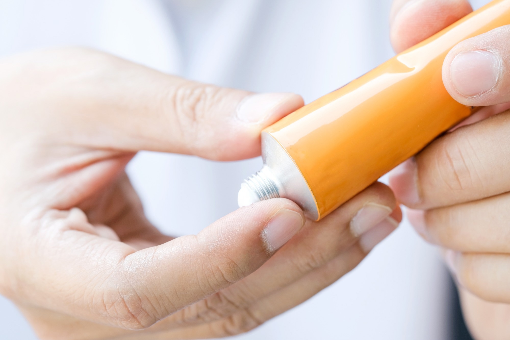
A recent study published in the journal Nature examines the use of the skin-permeable molecule poly[2-(N-oxide-N,N-dimethylamino)ethyl methacrylate] (OP) as a delivery system for insulin, a key drug in the treatment of type 1 and many cases of type 2 diabetes mellitus.
Small-molecule drugs are often designed to be absorbed through the skin. However, this has not been found feasible for large biomolecules, such as proteins and peptides like insulin.
Barriers to insulin delivery
Insulin is typically administered by intradermal injection. This method is painful, can induce fear of needles, and skin complications, all of which are linked to poor patient compliance. No successful noninvasive technique of insulin delivery has yet been reported.
Transdermal drug delivery offers several advantages, including improved patient compliance, convenience, increased active drug concentration by avoiding denaturation, and reduced first-pass metabolism of the drug. Among the greatest challenges in this approach is getting past the stratum corneum (SC) of the skin.
The SC is composed of dead and dried-out corneocytes surrounded by a well-ordered fatty matrix. Along with the epithelial tight junctions in the epidermis and dermis, this presents a barrier to drug penetration. Potential approaches include chemical penetration enhancers, electrically-driven devices that force the drug to penetrate the skin, and injection by ultrasound or jet rather than hypodermic needles, as well as microneedles. Being invasive, these are, however, associated with a higher risk of infection.
Cationic peptides can sometimes pass through the skin, bound to organic acids in the sebum and stratum corneum. However, this binding immobilizes them in the SC, preventing their deeper diffusion. Their only route lies through the hair follicles and sweat glands, comprising <0.1 % of skin area, meaning penetration is inefficient.
This prompted the current exploration of the novel polymer OP. The extreme skin permeability of OP-I does not involve any change in skin lipid ordering or structure. Instead, molecular dynamics simulations revealed that OP-I was adsorbed by stratum corneum fats faster than native insulin, diffusing rapidly through the lipids to reach the dermis and subcutaneous tissue.
This was characterized by a transition from its protonated cationic state (at a pH of 5 or lower) to a zwitterion during its skin passage (at neutral pH). This pH-dependent charge shift aligns with the skin’s acidic-to-neutral gradient and is central to OP’s transport behavior.
This switch corresponds to the pH change of the skin layers, progressing from superficial to deep. On topical application to the skin, OP builds up in the acidic sebum, and in the fatty acids contained in the fat layering the cornified cells. In the deeper layers of the SC, which have a neutral pH, it becomes a polyzwitterion, thus favoring free diffusion by reducing electrostatic interactions with the stratum corneum lipids.
OP can thus pass quickly and smoothly through the skin into the blood and lymph vessels. OP and OP–insulin primarily enter systemic circulation through leaky lymphatic capillaries before reaching the bloodstream.
Study findings
OP diffusion
The researchers applied fluorescent-labeled OP to the skin surface of mice and minipigs (the skin of the latter being very similar to human skin) and tracked its passage through the skin using high-resolution imaging.
In mice, OP diffused throughout all skin layers within four hours of topical application, whereas the control polyethylene glycol (PEG) remained on the skin surface. In the epidermis and dermis, OP-I moved by membrane-mediated diffusion without entering the cell. This involved rapid “hopping” along adjacent cell membranes rather than intracellular transport.
Further confirmation was obtained by visualizing the OP-bound gold nanoparticles within the lipid lamellae of the intercorneocyte fatty layer. OP penetrates the skin with exceptional efficiency, entering the bloodstream within 30 minutes. Its concentration peaked approximately two hours later.
Insulin-conjugated OP
Recombinant human insulin was then conjugated to OP (OP-I), with pegylated insulin serving as a control, having a similar molecular mass of 5 kDa. OP-I had the same secondary structure as insulin. It also showed unchanged receptor binding and association-dissociation constants, indicating that it retained the receptor specificity and affinity of native insulin intact.
OP-I skin permeability was measured by the drop in blood glucose following topical application. When compared with unbound insulin, the conjugated insulin produced the same decrease in blood glucose.
Thus, the study suggests that OP-I behaves similarly to insulin, binding to the insulin receptor with unchanged specificity and activating downstream pathways that result in glucose-lowering effects. OP-I had a longer half-life than insulin, probably because of its zwitterionic nature that resists plasma protein binding and removal from the blood. This extension was modest (15 to 20 minutes vs. 5 to 10 minutes for native insulin).
Modeling of the skin permeation of OP-I over time using confocal laser-scanning microscopy (CLSM) showed its uniform spread throughout the epidermis in half an hour. In contrast, native insulin and PEG-I remained on the skin surface. Thus, OP-I had the highest permeability coefficient among the three, approximately 4.5 and 9-fold that of the PEG-I and insulin, respectively.
OP effects on insulin and blood glucose
OP-I achieved comparable plasma levels to subcutaneous insulin within two hours. After this point, its levels were 60 % to 600 % higher than with insulin. The other two molecules did not affect blood insulin levels.
In mice with type 1 diabetes, OP-I normalized blood glucose levels. Once in the blood, OP-I was taken up mainly by the liver, lungs and kidneys, inducing insulin activity. Its activity was prolonged compared to subcutaneous native insulin, which was rapidly cleared from the bloodstream without significant accumulation in these tissues. OP-I regulated blood glucose levels better in diabetic mice than other treatments.
Similar findings were observed in minipigs, with OP-I entering the dermis and subcutaneous tissue at four hours following topical application. It induced normal blood glucose levels within two hours and maintained them for 12 hours.
Notably, topical OP-I application did not irritate the skin or induce inflammation. Repeated application in both mice and minipigs caused no structural changes to the stratum corneum or signs of immune activation.
Toward needle-free insulin
The skin-permeable polymer may enable non-invasive transdermal delivery of insulin, relieving patients with diabetes from subcutaneous injections and potentially facilitating patient-friendly use of other protein- and peptide-based therapeutics through transdermal delivery.
Download your PDF copy now!
 Can ketogenic diets help PCOS? New analysis points to weight and insulin gains