Research reveals how invisible nanoparticles manipulate cellular messengers, undermining your gut’s delicate microbiome, raising new questions about the unseen health risks of environmental nanoplastics.
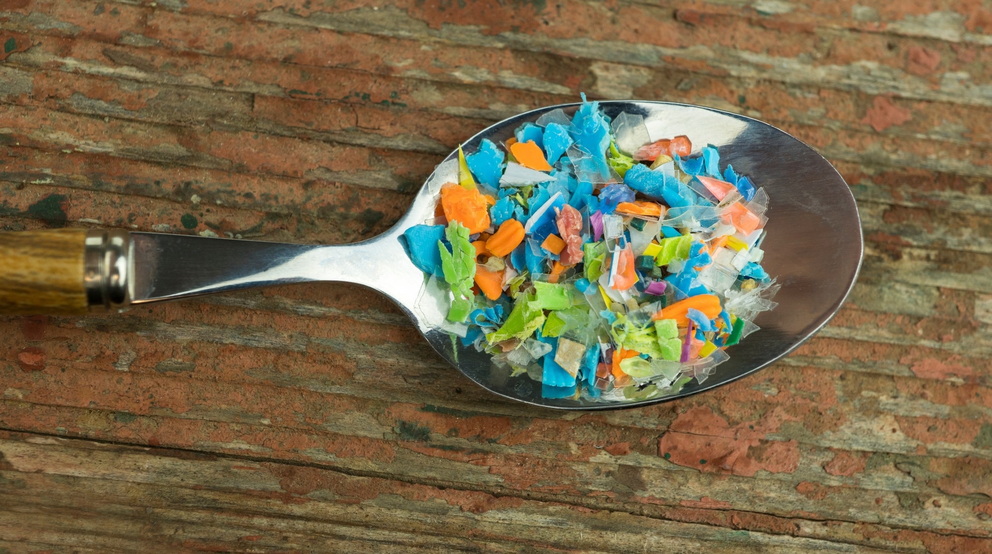
 Study: Polystyrene nanoplastics disrupt the intestinal microenvironment by altering bacteria-host interactions through extracellular vesicle-delivered microRNAs. Image credit: SIVStockStudio/Shutterstock.com
Polystyrene nanoplastics exposure may disrupt gut health by altering bacterial-host interactions and disturbing the intestinal microenvironment. A recent study published in Nature Communications investigated how polystyrene nanoplastic exposure affects human health, focusing on bacterial-host interactions.
The effect of nanoplastic exposure on human health
Humans are frequently exposed to plastic fragments throughout the food chain, which raises questions about their impact on the gut microbiome. The degradation of different types of plastics, such as polystyrene (PS), polyvinyl chloride (PVC), and polyethylene (PE), results in the development of microplastics (MP) and nanoplastics (NP).
Multiple studies have indicated that MP or NP exposure may cause hematopoietic damage, liver injury, and testicular disorders in mammals through gut dysbiosis. These studies have also shown that PS-MP and PE-MP exposure induce inflammation, immune imbalances, and gut barrier dysfunction. More specifically, PE-MP exposure alters gut microbial composition by favoring a selective increase of pathogenic Staphylococcus aureus. This NP also promotes intestinal inflammation.
Despite understanding the toxic effects of MP and NP in humans, few studies have explored the interaction between microscopic plastics, gut microbiota, and the host. Furthermore, the underlying mechanism by which microscopic plastic affects human health remains relatively under-researched.
Several studies have proposed that NPs are more harmful than MPs due to their smaller size. This allows them to penetrate tissues and organs, easily affecting their biological functions. Understanding the precise pathway through which NPs cause gut dysbiosis and affect intestinal health is essential.
Extracellular vesicles (EVs) are tiny, membrane-bound lipid bilayer sacs released by animal cells and bacteria. These spherical structures carry diverse contents, including DNA, RNAs, proteins, and lipids. EVs play a crucial role in intercellular communication. Previous studies have indicated that EVs often mediate the interaction between microbiota and the intestinal epithelium, influencing gut health and function.
About the study
The current study hypothesized that NP directly or indirectly impacts the microbiota composition through EVs. Several in vivo and in vitro experiments were performed to test this hypothesis. For instance, the size and number of NP used in this study were confirmed using nanoparticle tracking analysis (NTA).
Six-week-old male mice were exposed to fluorescently labeled NPs to study their distribution in organs. Cellular uptake of NPs, serum biochemical analysis, real-time PCR, and western blot were performed.
To understand how NP affects gut microbiota, microscopic polystyrene (100 nm) was orally administered to the mice four times a week for 12 weeks, particularly on days 1, 3, 5, and 7 of each week. A set of control mice, which were not treated with NP, was maintained for reference.
Study findings
NP (100 nm) accumulation was observed at varied time points ranging between 3 minutes and 48 hours. The current study detected significant levels of NP in the small intestine, liver, cecum, and colon of the study mice.
Oral NP exposure increased body weight compared to mice in the control group. However, the increase was moderate and not associated with significant changes in white adipose tissue or liver weight. No significant changes in liver weight or white adipose tissue weight were observed. Intestinal shortening was not observed in NP-exposed mice, implying that intestinal bacteria, not inflammation, was the primary target of NP-induced effects.
Biochemical analysis revealed that 12 weeks of NP exposure did not significantly modify serum aspartate aminotransferase (AST), alanine aminotransferase (ALT), creatinine (CRE), or blood urea nitrogen (BUN) levels. This finding suggests that the intestinal microbiota and barrier may be directly affected by NP.
The current study observed that NP could penetrate the enterocyte-like differentiated Caco-2 cells and mouse intestine after 24 hours of treatment. After entering, it reduces the expression of tight junction proteins, including zonula occludens-1 (ZO-1) and occludins (OCC). This disruption causes characteristic intestinal damage, including increased intestinal permeability or leaky gut.
Gene ontology (GO) analysis indicated that NP exposure significantly altered mice's intestinal gene expression and metabolic functions. Principal component analysis (PCA) of microRNA (miRNA) diversity in mouse feces revealed that NP exposure substantially modified miRNA profiles and reduced the diversity of specific miRNAs. Further in-depth analysis uncovered the role of miRNAs as a regulator of primary physiological functions, particularly those associated with intestinal cell junctions.
Experimental findings suggested that NP could interfere with tight junction protein expression by regulating miRNAs in intestinal cells, ultimately disrupting the intestinal environment. Predictive analysis indicated NP exposure upregulates miRNAs, such as as-miR-98-3p, has-miR-548h-3p, has-miR-548z, has-miR-548d-3p, has-miR-548az-5p, has-miR-12136, and has-miR-101-3p, which affects the ZO-1 gene expression.
Additionally, the study identified that NP exposure increased the expression of mouse-specific miRNAs, such as mmu-miR-501-3p and mmu-miR-700-5p, which also interfere with ZO-1 and MUC-13 expression.
Immunocytochemistry (ICC), qPCR, and Western blot analysis revealed that NP treatment reduces MUC-13 expression in mice and enterocyte-like differentiated Caco-2 cells.
With prolonged NP exposure, unique bacterial species initially increased and decreased. The most notable effect was a shift in the relative abundance of specific bacterial taxa rather than a simple loss of overall diversity. For instance, Lactobacillaceae decreased, and Ruminococcaceae increased.
The study also noted that Akkermansia, a beneficial next-generation probiotic bacterium, increased abundance in NP-exposed mice, particularly at later times. Experimental findings demonstrated that the impact of NP on the gut microbiome was not directly caused by NP toxicity but via other mechanisms.
Specifically, the study shows that the changes were mediated through extracellular vesicles (EVs) derived from intestinal cells and certain bacteria rather than the direct toxic effects of NP on bacterial growth. Lachnospiraceae sp.-derived EVs did not influence the growth of intestinal bacteria.
The novelty of this study lies in uncovering a specific mechanism. NP alters the gut microenvironment by modulating EV-mediated delivery of miRNAs, which then disrupt the intestinal barrier and selectively influence the growth of bacterial taxa. This represents a newly described pathway in the context of NP toxicity.
Conclusions
The current study suggested that NP impacts specific bacterial taxa, including Lachnospiraceae and Ruminococcaceae. The alteration in the gut microbiome upon NP exposure is mediated by host-microbiota interactions through EV. NP ingested by Lachnospiraceae triggered suppressed mucin-13 expression.
Additionally, EVs released from goblet-like cells after NP exposure promoted the growth of Ruminococcaceae, highlighting a complex interplay between host-derived and bacterial-derived vesicles.
Further research on the impact of NP on human and environmental health is needed. While these findings provide new insights into how NP may disrupt gut health, it is essential to note that the experiments were conducted in mice. The relevance of the doses and findings to typical human exposures remains to be determined.
Download your PDF copy now!
Journal reference:
- Hsu, W. et al. (2025) Polystyrene nanoplastics disrupt the intestinal microenvironment by altering bacteria-host interactions through extracellular vesicle-delivered microRNAs. Nature Communications. 16(1), 1-13. https://doi.org/10.1038/s41467-025-59884-y https://www.nature.com/articles/s41467-025-59884-y