The use of biospecimens is common in research, from initial research stages to late-phase clinical trials. As a result, a comprehensive understanding of pre-analytical deviations in collection, processing, and storage is vital to downstream applications and data analysis. In this article, processing and its effect on specimens is discussed.
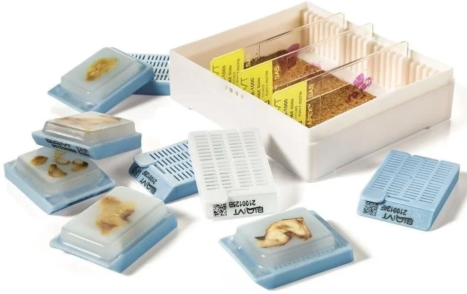
Image Credit: BioIVT
Types of fixation used in research
The two main fixation formats used by researchers are formalin fixation followed by paraffin embedding (FFPE) or snap-frozen in liquid nitrogen (referred to as Fresh Frozen or FF).
There is less of a demand for fixation in reagents other than formalin or to embed frozen specimens in OCT (a standard freezing media). The use of RNA stabilization reagents, such as QIAGEN’s RNAlater®, has also become more prominent in research.
RNAlater is specifically designed for use with cells and tissues. It rapidly permeates tissue, stabilizing and protecting cellular RNA for downstream gene expression analysis.
Can you switch between fixation types?
A fresh frozen sample may be embedded in OCT. This enables the sample to be sectioned on a cryostat, producing better slides for downstream staining applications.
However, converting an FF sample to FFPE results in a suboptimal tissue block. This is because the process of snap-freezing a specimen converts water into ice, causing a “freezing artifact,” which is usually detrimental to tissue morphology.
Considerations for FF fixation
In cases where FF is necessary, steps are taken to limit the freeze artifact using special reagents and tools. Due to the labor-intensive nature of these steps, they are only implemented when necessary.
There are also certain tissues with higher water content, such as the brain, fatty tissue, lymph nodes, and tissue containing a lot of blood, which produce worse freeze artifact effects.
Important factors for the fixation process
A major factor influencing the fixation process is tissue section size. For both FFPE and FF procedures, the diffusion rate of formalin perfusion or the freezing process limits the maximum tissue size for acceptable results. Generally, a limit of 3 mm thickness is implemented for both processes. Of less importance is surface area, as diffusion occurs through the cross-section of the tissue.
Size also affects the processing time of samples, with smaller tissue requiring less processing processing. The type of tissue must also be considered and adjusted based on fat and bone content, for example.
Sample storage
Due to their small size, fixated tissues could easily be stored in vials or tubes. However, it is not recommended, particularly if staining is planned for downstream analysis. This is mainly because storage and removal of tissue from a tube or vial damages the sample.
Often vials are not the correct size to fit the tissue, resulting in sample deformation and preventing downstream histological visibility of the tissue layers and morphology. This is particularly important for tissues such as the skin, intestine, and endometrium.
On top of this, mechanical manipulation activates stress genes, altering downstream analysis of gene expression.
In cases where tissue cannot be removed from a vial, the container must be cut or crushed resulting in tissue damage, thereby causing fragmentation and destruction of the tissue block.
If storage in a vial or tube is unavoidable, the use of a certified cryogenic vial is required, rather than an Eppendorf tube or other container unsuitable for cryogenic storage.
For the best results, histology cassettes are recommended for storage of FFPE and FF samples. Careful placement of the tissue in a cassette enables decreased variation and additional artifacts in the tissues.
Sectioning FF tissues
As OCT can interfere with downstream processing, tissues are not usually embedded in OCT. To section a non-OCT-embedded tissue block, DEPC-treated RNA-free water is used to adhere the tissue section directly to the chuck, which enables the creation of sections from the tissue.
DEPC is used to create RNase-free water, offering a solution that limits any effects on downstream analysis. The water is autoclaved after DEPC treatment, which inactivates the DEPC by causing hydrolysis of diethylopyrocarbonate, resulting in the release of CO2 and EtOH. Therefore, based on a half-life of 30 minutes, autoclaving 1 L of DEPC-treated water for 15 minutes results in DEPC-free water.
About BioIVT
BioIVT, formerly BioreclamationIVT, is a leading global provider of high-quality biological specimens and value-added services. We specialize in control and disease state samples including human and animal tissues, cell products, blood, and other biofluids. Our unmatched portfolio of clinical specimens directly supports precision medicine research and the effort to improve patient outcomes by coupling comprehensive clinical data with donor samples.
Our Research Services team works collaboratively with clients to provide in vitro hepatic modeling solutions. And as the world’s premier supplier of ADME-Tox model systems, including hepatocytes and subcellular fractions, BioIVT enables scientists to better understand the pharmacokinetics and drug metabolism of newly discovered compounds and the effects on disease processes. By combining our technical expertise, exceptional customer service, and unparalleled access to biological specimens, BioIVT serves the research community as a trusted partner in ELEVATING SCIENCE®.
Sponsored Content Policy: News-Medical.net publishes articles and related content that may be derived from sources where we have existing commercial relationships, provided such content adds value to the core editorial ethos of News-Medical.Net which is to educate and inform site visitors interested in medical research, science, medical devices and treatments.