Biophotonics, the melding of biology, medicine, artificial intelligence (AI), and photonics, is fast becoming a transformative approach for analyzing biological systems. By using light to interrogate biological materials, biophotonics enables both highly sensitive and minimally invasive measurements across fundamental research, diagnostics, and therapeutic monitoring.1
The South-Central region of the United States occupies a keystone of research in medical biophotonics and AI, with internationally recognized programs at the University of Texas at San Antonio (UTSA) and the University of Houston (UH). This year, with Pittcon taking place in San Antonio, the conference is situated directly within this growing ecosystem, creating a natural intersection between global analytical science and regional biomedical innovation.
Biophotonic technology and artificial intelligence
The field of biophotonics can be split into three overlapping domains:
- Bioimaging: Used to characterize biological specimens across spatial scales, from nanoscopic intracellular structures to whole tissues. Molecular composition, electronic chromophores, structural proteins, refractive index changes, and phase boundaries can be resolved through an array of techniques, including fluorescence imaging, optical coherence tomography (OCT), photoacoustic imaging, Raman scattering, and Brillouin scattering.
- Biosensing: Photonic approaches detect biomolecules, including disease-specific biomarkers, with sensitivities reaching molecular or even single-molecule levels. These capabilities enable early disease detection and detailed molecular profiling.
- Treatment and treatment control: Light sources are employed for precise and minimally invasive interventions, while imaging and sensing methods provide real-time feedback on treatment efficacy and recovery.
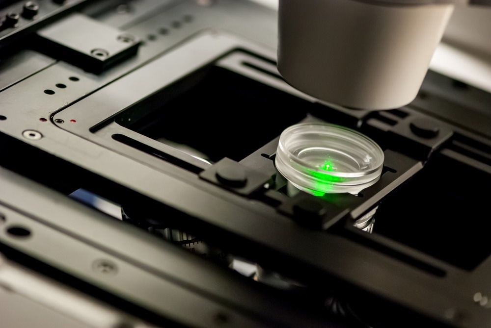 Image Credit: Micha Weber/Shutterstock.com
Light-based methods are advantageous due to the kind of measurements they support. By allowing non-contact and non-toxic observation of living systems, they facilitate rapid acquisition of real-time data, enable highly sensitive detection, and make it possible to follow biological processes across a wide range of timescales, from fast molecular events to slower physiological changes.
The integration of AI makes these measurements interpretable at scale. Modern machine learning methods, including deep learning, can analyze large spectral and imaging datasets that would be impractical to assess manually. This improves diagnostic accuracy, enhances image quality, supports real-time decision-making, and provides more individualized treatment strategies.
At the same time, clinical use requires that these systems be transparent and interpretable. Explainable AI, high-quality training data, and collaboration between photonics researchers and data scientists are therefore necessary to ensure that AI-driven outputs can be validated before integration into medical workflows.
Together, biophotonics and AI are transforming medicine into earlier and more personalized forms of care, enabling continuous physiological monitoring, early disease detection, and real-time guidance of therapy. In oncology, this is exemplified by image-guided interventions and photodynamic treatments; in broader healthcare, it takes the form of patient-specific measurement and interpretation, rather than relying on sparse or indirect clinical signals.1
Next-Generation Raman Spectroscopy Utilizing AI
Raman spectroscopy is a notable example of how a biophotonic technique can transition from the laboratory to clinical relevance. It provides label-free molecular fingerprints based on the intrinsic vibrational signatures of biomolecules, capturing information about chemical composition and the local environment.1
In practice, Raman methods have been used to monitor bile reflux through bilirubin absorption in fiber-optic catheter systems and to identify microorganisms based on characteristic spectral patterns. Surface-enhanced Raman scattering, utilizing nanostructured gold or silver substrates, can amplify otherwise weak signals by several orders of magnitude, thereby extending detection limits into regimes relevant for clinical and microbiological analysis.1
For this technology, difficulty lies less in generating data and more in interpreting it. Differences between biological states can be subtle, and spectral fingerprints often vary only slightly with condition. AI provides a way to convert this complexity into usable information by classifying spectra, extracting patterns, quantifying components, and allowing the detection of organisms or biomarkers that would be difficult to recognize by conventional analysis.1
At Pittcon, this translational trajectory is the focus of the symposium talk: "Next-Generation Raman Spectroscopy Utilizing AI: From High-Throughput Platforms to Endoscopic Probes for Clinical Translation" by Professor Juergen Popp of the Leibniz Institute of Photonic Technology.
Professor Popp’s presentation describes a trifecta of translational Raman approaches from a high-throughput Raman blood analysis platform and the Raman invaScope, to multimodal endoscopic probes that extend Raman from point measurements to morphochemical imaging. Together, these approaches demonstrate how Raman can evolve from a research tool into a real-time clinical decision technology.
Plasmonic technologies and infectious disease testing
Infectious disease diagnostics provide a clear example of where these technologies are particularly important. PCR and lateral flow assays are now routine, but they remain limited in their ability to detect multiple targets simultaneously and, in the case of lateral flow tests, often in terms of sensitivity and reliability.2,3
Plasmonic biosensors offer a different approach. Methods such as surface plasmon resonance, localized plasmon resonance, plasmonic fluorescence enhancement, and surface-enhanced Raman scattering enable label-free detection of multiple biomarkers in a single measurement. These techniques have already been applied to diseases such as tuberculosis, malaria, and Lyme disease.4
This is particularly relevant in point-of-care settings, where efficiency matters as much as analytical performance. Traditional methods, such as ELISA and PCR, depend on laboratory infrastructure and trained personnel, whereas plasmonic platforms can be adapted into compact formats that meet the WHO’s ASSURED criteria, making them more practical in resource-limited environments.4
This theme is explored in the Pittcon talk Plasmonic-enhanced Fluorescence for Rapid, Multiplexed Diagnostics by Professor Nathaniel Cady of the University at Albany. Prof. Cady will expound the grating-coupled fluorescent plasmonics (GC-FP) biosensor platform, which has enabled multiplexed antibody and antigen detection with a quantitative, linear response. The platform supports multiple testing formats, from microfluidic systems to point-of-care devices, and extends to nucleic acid hybridization and protein interaction assays.
Conclusion: Translational biophotonics at Pittcon
Taking place in Texas, Pittcon is situated in a region where biophotonics and AI are becoming a significant focus of research. The conference provides a setting in which ideas developed in academic and technical contexts can be considered in terms of their practical implications for medicine.
Hamamatsu Corporation, which is exhibiting at Booth 3337, exemplifies the instrumentation backbone that supports this progress, supplying photonic components and detectors that underpin imaging, sensing, and spectroscopy platforms.
More information on sessions, speakers, and exhibitions is available at pittcon.org, where attendees can explore how biophotonics and AI are becoming integral to modern bioanalysis and medicine.
References and further reading
- Baldini, F., et al. (2025). Shining a Light on the Future of Biophotonics. Journal of Biophotonics. DOI: 10.1002/jbio.202500148. https://onlinelibrary.wiley.com/doi/full/10.1002/jbio.202500148.
- Khatmi, G. et al. (2024). Lateral flow assay sensitivity and signal enhancement via laser µ-machined constrains in nitrocellulose membrane. Scientific Reports, 14(1). DOI: 10.1038/s41598-024-74407-3. https://www.nature.com/articles/s41598-024-74407-3.
- Tolle, H., et al. (2025). Implementation of Point-of-Care PCR-testing for the diagnosis of respiratory infections in vulnerable patient populations. PubMed, (online) 20(7), pp.e0307621–e0307621. DOI: 10.1371/journal.pone.0307621. https://journals.plos.org/plosone/article?id=10.1371/journal.pone.0307621.
- Li, Z., et al. (2019). Plasmonic-based platforms for diagnosis of infectious diseases at the point-of-care. Biotechnology Advances, 37(8), pp.107440–107440. DOI: 10.1016/j.biotechadv.2019.107440. https://www.sciencedirect.com/science/article/abs/pii/S0734975019301405?via%3Dihub.
About Pittcon
Pittcon is North America’s largest conference and exposition in laboratory science, attracting thousands of industry, academia, and government attendees and exhibitors from around the world in. Pittcon goes beyond the typical conference; it's a global community dedicated to advancing science through education, collaboration, and philanthropy. By bringing together scientists, researchers, educators, students, and industry leaders, Pittcon creates opportunities to learn, connect, and inspire innovation. Beyond the event itself, more than 90% of Pittcon's net proceeds support science education and outreach initiatives locally and globally, funding scholarships, grants, STEM programs, laboratory improvements, and other efforts that strengthen communities and expand access to science.
Pittcon delivers world-class educational and professional development opportunities through its Technical Program, Short Courses, and Networking Workshops and Roundtables. Attendees can explore the latest scientific research through symposia, oral and poster presentations, enhance their skills with expert-led deep-dive courses, and connect with peers through interactive workshops and networking experiences designed to foster collaboration and career growth.
The Pittcon Expo brings the scientific community together in an immersive environment where attendees can discover the latest laboratory technologies, meet with leading instrument and service providers, and experience science in action. Beyond the exhibit booths, the Expo Floor features engaging attractions, educational experiences, demonstrations, and networking spaces, while receptions, parties, and social events create opportunities to build lasting connections with colleagues from around the world.
Sponsored Content Policy: News-Medical.net publishes articles and related content that may be derived from sources where we have existing commercial relationships, provided such content adds value to the core editorial ethos of News-Medical.net, which is to educate and inform site visitors interested in medical research, science, medical devices and treatments.