Bioprinting is increasingly recognized as a potentially revolutionary technology in cancer research, offering new opportunities for drug screening, disease modeling, and personalized medicine.
Bioprinting fabricates complex, multicellular 3D tissues that closely mimic the tumor microenvironment, allowing researchers to study cancer with increased accuracy and reproducibility.
This article outlines bioprinting’s unique value proposition in cancer modeling, highlighting how this technology can accelerate progress toward more effective therapies and address the limitations of traditional methods.
Cancer research has historically relied on two-dimensional cell cultures and animal models. These systems have offered valuable insights, but they tend to fall short in replicating the complexity of human tumors. For instance, two-dimensional monolayers lack the cell heterogeneity, spatial architecture, and dynamic interactions found in actual tumors.
Animal models offer the benefit of studying cancer within a full biological system, but these approaches also face numerous challenges.
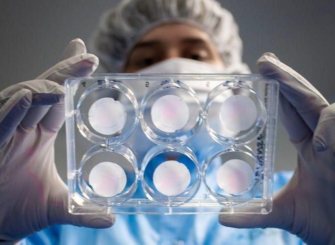
Image Credit: Poietis
Cancer is a multifactorial disease that can be affected by a range of environmental, genetic, and systemic parameters. This complexity typically introduces too many variables in animal models, meaning it is difficult to isolate the specific mechanisms or causes of treatment response or disease progression. Findings can, therefore, be difficult to interpret and less translatable to the human context.
Bioprinting, contrastingly, is a powerful tool able to simplify this complexity. Precisely controlling the structural, cellular, and extracellular components of a tumor model allows researchers to focus on specific variables in a highly reproducible and human-relevant environment: for example, stromal interactions, the role of immune cells, or hypoxia.
The ability to isolate and manipulate specific features of the tumor microenvironment offers clearer insights into drug effects and cancer biology.
Key benefits of bioprinting in cancer modeling
Improved physiological relevance
Bioprinted cancer models better preserve tumor biology’s critical features, including:
- Cell-cell and cell-matrix interactions
- Gradients of nutrients, oxygen, and drugs
- Tissue mechanical cues and stiffness
These factors impact metastasis, tumor progression, and treatment response, making bioprinted models more predictive of in vivo behavior than traditional platforms.
Personalized tumor models
Researchers can bioprint personalized cancer models using patient-derived cells; these models more closely reflect the specific characteristics of a patient’s tumor. This enables personalized drug screening and treatment selection, improves clinical outcomes, and reduces trial and error in therapy.
High-throughput drug screening
Bioprinting enables the automated fabrication of miniaturized tumor models in multi-well formats. These models are ideal for high-throughput screening, supporting the rapid and cost-effective testing of a diverse array of therapeutic agents, including targeted therapies, chemotherapies, and immunotherapies.
Integration of tumor microenvironment (TME) components
The capacity to co-print cancer cells with endothelial cells, fibroblasts, and immune cells offers researchers a powerful tool to study the TME’s impact on tumor treatment resistance and biology. This capability makes bioprinted models extremely valuable for immuno-oncology and stromal targeting strategies.
An alternative or complement to animal models
Animal models remain useful in understanding systemic responses, but bioprinting complements these models by offering a reductionist approach that is still relevant for humans.
Economic and ethical benefits
Bioprinting's reduced reliance on animal models means that it represents a more ethical and typically more cost-effective alternative for preclinical testing. It also better aligns with the 3Rs principles (replacement, reduction, and refinement) for animal research.
Summary
As the boundaries of cancer research continue to expand, bioprinting is becoming increasingly recognized as a powerful way to balance complexity with control, shifting the paradigm beyond the constraints of conventional models.
It may seem reductionist to isolate components of the tumor microenvironment, but this modularity enables researchers to deconstruct multifactorial systems and better examine interactions with increased clarity.
Bioprinting enhances translational accuracy, enabling more predictive preclinical data by offering models that better reflect human physiology. This pioneering technology may help expedite regulatory clearance and accelerate the development of safer and more effective therapies.
About Scintica Instrumentation Inc.
At Scintica, we advance science and medicine by supplying researchers with reliable research instrumentation and equipment. Our carefully selected portfolio of imaging systems, research tools, and supporting technologies is designed to reduce complexity and help scientists focus on what matters most, generating
meaningful results.
We partner closely with the preclinical research community to connect teams with solutions that are scientifically robust and built to support research challenges. From system selection through long-term support, our goal is to make research more productive, efficient, and impactful.
Sponsored Content Policy: News-Medical.net publishes articles and related content that may be derived from sources where we have existing commercial relationships, provided such content adds value to the core editorial ethos of News-Medical.net, which is to educate and inform site visitors interested in medical research, science, medical devices and treatments.