Sponsored Content by Merck KGaAReviewed by Louis CastelOct 2 2026
Three-dimensional culture is gaining acceptance as a paradigm for cell cultivation due to its capacity to replicate the in vivo microenvironment of cells in three dimensions.1 This approach promotes improved cell viability, especially for primary cells, while allowing for easy standardization.2 The FDA is currently placing significant focus on this area.
Three-dimensional (3D) culture can be divided into suspended 3D culture and scaffold-based 3D culture.3,4The latter is a significant research topic, focusing on the development of sophisticated biomaterials. By embedding cells within these materials, phenotypes become more apparent and cell functions are enhanced.5
Biomaterials can be engineered for printability.6 Three-dimensional bioprinting technologies enable the customization of the shape and internal structure of these materials, while precisely positioning the cells in the materials in three dimensions. This allows for highly controllable 3D cell co-culture capable of simulating tissues and organs.7 Employing 3D bioprinting technology to build 3D cell co-culture systems provides a sophisticated model for drug screening thanks to its highly biomimetic nature.
This article looks at recent progress in 3D bioprinted tissue models and outlines 3D bioprinting developments in pharmaceutical discovery. Finally, it examines the future of 3D bioprinting-based drug discovery and how it compares to other biomimetic techniques such as organoids and organ chips.8,9
3D bioprinted tissue models
Drug screening based on three-dimensional bioprinting relies on 3D bioprinted tissue models, which are rapidly advancing, including 3D-bioprinted liver. The liver’s complex vascular network plays a crucial role in substance exchange during drug metabolism. Using 3D bioprinting to develop a vascularized in vitro liver model is an important goal.
A recent study by Kang and colleagues accomplished this by engineering a heterogeneous, multicellular, multi-material liver lobule array.10 The research team designed a pre-defined 1 mm structure that included a 150 μm diameter central vein channel, high-density liver cells with embedded endothelial cells, 10 μm resolution micro-nozzles, and an outer endothelial lining to form a luminal tube.
The scientists used a battery of tests to analyze formation status, structural integrity, mechanical performance, liver function expression, and amiodarone-induced liver toxicity. The bioprinted body yielded ideal outcomes in all assessments, signifying its potential as an in vitro liver construction model.
Three-dimensional coaxial printing technology has recently demonstrated significant potential for directly producing injectable hollow structures. Specifically, Singh et al. have achieved substantial progress by creating a decellularized kidney-derived bioink blended with a mixture of sodium alginate, renal proximal tubule epithelial cells (RPTECs), and human umbilical vein endothelial cells (HUVECs) for bioprinting kidney tubular structures.11
Their bioprinter uses a coaxial nozzle to concurrently extrude bioink containing free substances and cells. Following shell crosslinking, the middle component is removed to form complete single- or double-layer tubular structures. Furthermore, the RPTEC and HUVEC tubes have been successfully transplanted into the peritoneal area of immunodeficient mice and implanted into host kidney tissue.
Bioprinting multi-tubular tissue structures represents a promising approach. Crucially, it has applications in drug reabsorption and excretion, as well as in the replacement of damaged kidneys.
Deng and colleagues introduced a new approach for building hollow tubular structures. By utilizing a time-dependent crosslinking strategy, they successfully controlled tubular wall thickness. A major benefit of this technique is its compatibility with cell seeding, enabling the replication of various tubular structures relevant in vivo, including blood vessels and renal tubules.29
Faheem Ullah et al. successfully synthesized a new bioink, PEO-CS-PMMA, which was used to construct 3D-bioprinted skin tissue structures.12 The bioink was prepared via the copolymerization of PEO, chitosan, and PMMA to enhance its density, viscosity, and printability while preserving thermal stability. The resulting bioink was highly organized and porous, facilitating water absorption and supporting the release of growth factors and bioactive substances that encourage healing and recovery.
Ma and colleagues described a technique for cross-linking bioinks with Ca2+ to produce gelatin-alginate for a 3D-bioprinted hydrogel scaffold with sufficient stability and biocompatibility.13 The scaffold can simulate the physical microenvironment of dermis and subcutaneous tissue by encapsulating adipose stem cells to encourage their proliferation and migration.
The unique gradient composite scaffold supports angiogenic and wound-healing effects by improving paracrine secretion from adipose stem cells. By providing a continuous and unique structure for adipose stem cells, this 3D-printed scaffold promotes skin regeneration and can be expanded for use in complex tissue injury models.
Lee and colleagues employed fiber-optic-assisted bioprinting to cross-link methacrylate-based hydrogels (GelMA) and construct biofunctional cell-loaded structures by choosing adequate photocrosslinking process conditions, including printing temperature.14 Their study triggered C2C12 mouse myoblasts and human adipose stem cells (HASCs) to form cell-loaded structures for muscle regeneration.
Compared to conventional printing procedures, the cell-loaded structures demonstrated higher cellular alignment and myogenic activity. Models built with HASC also exhibited superior muscle regeneration compared to those constructed without topographical cues.
However, factors affecting cellular activity, such as cellular metabolism, continue to pose challenges, resulting in inadequate oxygen transport in the 3D cellular constructs for muscle tissue regeneration. Hwangbo and colleagues tackled this problem by introducing photosynthetic cyanobacteria (Alcaligenes longum) into Gelma bioink. Using in situ electronic bioprinting, they prepared cell-loaded scaffolds that encourage cellular arrangement and muscle formation.15
They determined that this strategy could serve as an effective therapy for severe skeletal muscle defects by analyzing the combined effects of bioactive components in bioinks and bioprinting that concurrently support electronic fields. Bionic spinal scaffolds, based on 3D bioprinting technology, can effectively simulate both the shape and structure of spinal cord tissue and foster the healing of spinal cord injuries. However, they have not yet succeeded in simulating the biological functions of the spinal cord because of inadequate electrical conductivity.
Gao and colleagues engineered a novel conductive hydrogel, PEDOT:LS, which was used in a bionic scaffold based on GelMA, hyaluronic acid methacrylate (HAMA), and poly (3,4-ethylene dioxythiophene) to simulate the electrical conductivity of the spinal cord.16
The conductive hydrogels exhibited mechanical characteristics comparable to those of natural spinal cord tissue, and neural stem cell (NSC) culture demonstrated high survival rates. Relative to non-conductive scaffolds, the 3D bioprinting-based conductive scaffolds substantially promoted neuronal differentiation of NSCs in vitro and enhanced hind limb motor function recovery in a rat spinal cord complete transection model.
In a study by Zhang and colleagues, inorganic calcium silicate (CS) nanowires were introduced into bioink for innervated bone regeneration using 3D bioprinting technology.17 The researchers developed a model that combines nerve and bone-associated cells printed in an orderly fashion and encapsulated by CS nanowires. These nanowires improve long-term viability and proliferation of encapsulated cells while promoting osteogenesis and neural differentiation.
By incorporating inorganic nanomaterials into bioinks and 3D bioprinting technology, this study presents a promising approach for regenerating complex bone tissue.
Combining 3D bioprinting technology, fused deposition modeling (FDM) technology, and light-cured additive manufacturing (DIW) has led to significant advancements in cartilage and bone tissue engineering. Chen and colleagues18 prepared a biphasic bone scaffold using alginate-gelatin hydrogel (A-G) as the bioink and polycaprolactone (PCL) to enhance mechanical stability and general performance.
The integration of hydroxyapatite into the PCL improved the bone phase of the scaffold, resulting in enhanced bioactivity. Importantly, the physical and biological analysis of the scaffold in both the bone and cartilage phases validated its sound biological effects in both the short and long term, demonstrating its potential as an interface material between cartilage and bone.
3D bioprinting for drug screening
In contrast to 3D bioprinting-based tissue models, 3D bioprinting-based drug screening remains in its early stages. Hong and colleagues19 applied extrusion-based 3D bioprinting technology to produce a gelatin-sodium alginate-based construct embedding MCF-7 cells and observed that the MCF-7 cells auto-aggregate into spheroids.
These spheroids maintained their CD44 high /CD24 low /ALDH1 high drug-resistant phenotype and demonstrated elevated expression levels of drug resistance markers, including the GRP78 chaperon and ABCG2 transporter. This superior resistance was validated using camptothecin and paclitaxel.
Li and colleagues fabricated a 3D-printed breast cancer model using a hydroxyethyl cellulose/alginate/gelatin (HCSG) composite biomaterial.20 They first demonstrated the potential of 3D bioprinting technology in a structure–activity relationship study by examining the pharmacodynamics of 13 amino acid-based flavone phosphoramidates. Three-dimensional printed models exhibited distinct pharmacological activity properties compared to 2D monolayer models (Figure 1).
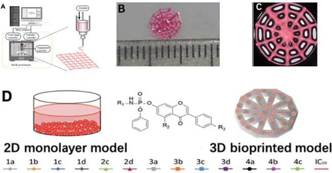
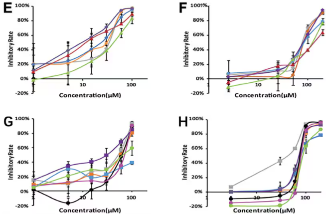
Figure 1. 3D printed breast cancer model. A) Workflow diagram. B-C) Schematic diagram of 3D printed “Spider web” HCSG breast cancer model. D) Investigating pharmacodynamics of 13 amino acid-based flavone phosphoramidates with 2D monolayer model and 3D printed model. Image Credit: © 2020, Zhejiang University Press.
Qiong Liu et al. created tubular structures within the GelMA gel using sacrificial extrusion-based 3D bioprinting technology.21 Cholangiocarcinoma (CCA) cells were allowed to attach to the surface of the tubular structure to mimic human biliary tract cancer. In this 3D printing model, CCA was overgrowing in a thickening manner, producing bile duct stenosis, which was expected to be analogous to the in vivo configuration.
CCA cells demonstrated greater sensitivity to anti-tumor therapeutics than traditional 2D cell models, providing a new pharmaceutical screening model for bile duct cancer treatment. To increase biocompatibility, decellularized extracellular matrix (DECM) is frequently incorporated into printable biomaterials.22
Janani et al. created two different bioinks based on tissue-specific DECM of liver cells.23 These bioinks exhibited exceptional printability and rheological characteristics while supporting the printing of both parenchymal and non-parenchymal cells of liver lobules.
The print model demonstrated dose-dependent clinical liver toxicity reactions to acetaminophen and troglitazone, offering a robust platform for liver toxicity screening. Human-induced pluripotent stem cells (hiPSCs) are a source of cells capable of differentiating into normal human cells.24 When used with 3D bioprinting technology, these human cells enable the replication of human tissues or organs.
He Jianyu et al. constructed a human liver model by printing hiPSC-induced-hepatocytes.25 Compared to conventional 2D culture models, hiPSC-induced hepatocytes in this model exhibited improved mRNA expression related to human liver-specific functions, as well as acetaminophen-induced liver toxicity.
Rency Geevarghese et al. created a bioink composed of multiple components, including alginate, diethylaminoethylcellulose, gelatin, and collagen peptides, and conducted experimental evaluations to ensure its suitability for 3D model construction.26
To evaluate the efficacy of their models, the team examined the growth and proliferation of A549 cells encapsulated within 3D-bioprinted structures. They demonstrated that 3D bioprinting-based tissue models offer a more effective platform for drug screening by simulating native tissue characteristics and serving as a superior alternative to conventional 2D cell toxicity assessment.
The scientists qualitatively and quantitatively confirmed the potential of their 3D-bioprinted structures as drug screening models using apoptosis assays and MTT assays. In conclusion, their findings highlight the potential of 3D bioprinting to transform drug screening practices by offering a more reliable and effective means for assessing therapeutic effectiveness.
Zicheng Fan et al. employed dot extrusion printing (DEP) technology to generate precise positioning and adjustable size of hepatocyte-laden gelatin methacryloyl (GelMA) hydrogel microbeads and HUVEC cell-containing gelatin microbeads, constructing endothelialized liver lobule-like structures.27
Their experimental findings showed that the appropriate GelMA concentration simulated the naturally occurring microenvironment, enhancing hepatocyte growth and propagation while offering a suitable surface for tight junctions and endothelial cell growth and proliferation.
Compared to 2D hepatocellular carcinoma cell models, the model generated using DEP technology also improved the efficacy of the antitumor therapeutic sorafenib. The endothelial barrier formed by HUVEC prevented the spread of sorafenib. The authors determined that DEP technology has potential for constructing 3D stromal and cancer cell models to support their growth and multiplication, creating a complex tumor microenvironment.
The endothelialized hepatocellular carcinoma models based on 3D bioprinted liver lobule-like structures can mimic pharmacodynamics similar to real-life conditions, making them indispensable tools for therapeutic testing.
Xue Liu et al. established a reproducible technique for inducing endothelial cells of different complexity by 3D bioprinting human keratinocytes, fibroblasts, pericytes, and multifunctional stem cells to construct skin models of varying physiological complexity.28 These included human epidermis, non-vascularized, and vascularized models used for high-throughput drug screening on multiwell plates.
This innovative combination of 3D cell culture and physiologically relevant 3D tissue models enabled high-throughput pharmacological research for the first time. The model was also evaluated for the efficacy of potential therapeutics for AD, with the findings correlating with clinical treatment data, underscoring the model’s value in 3D tissue disease modeling and pharmaceutical screening. This research serves as a valuable tool for future pharmacological research on multiwell plates and further contributes to 3D tissue disease modeling.
Perspectives
In addition to 3D bioprinting, organ-on-a-chip and organoids are two other biomimetic methods that can be utilized in pharmaceutical discovery.
Organ-on-a-chip technology uses micro-fabricated cell culture systems to precisely simulate the structure, function, and microenvironment of human organs. These microdevices consist of chambers, microfluidic channels, and sensors capable of simulating the intricate physiological environment of organs including the heart, liver, lung, kidney, and brain.
Essentially, the chip simulates blood circulation, cell metabolic activity, and the mechanical forces that act on the organ. Organ-on-a-chip technology offers multiple advantages over conventional cell culture and animal models.
First, it can recreate complex organ microenvironments, including the physical and chemical cues that govern cell growth and function. Second, it permits instantaneous monitoring and evaluation of cellular and molecular changes, allowing scientists to observe drug responses and disease progression directly. Third, it provides a more ethical and affordable alternative to animal testing, while offering the potential for personalized medicine through patient-derived cells.
Organoids are 3D structures that closely resemble the gross and microscopic anatomy of an organ or tissue. They are constructed from self-organizing stem cells that differentiate to form various types of cells found in the desired organ, giving rise to intricate functional tissues.
Various stem cell types can be used to create organoids, including embryonic stem cells, induced pluripotent stem cells, and adult stem cells. Organoids are indispensable instruments in pharmaceutical development, disease modeling, regenerative medicine, and personalized medicine. They offer substantial advantages over conventional two-dimensional cell cultures and animal models in human disease research and drug testing.
By accurately representing the actual organ, including the microenvironment and tissue architecture, organoids enable more precise and dependable pharmaceutical screening.
In addition, they reduce the need for animal testing and offer a robust instrument for elucidating the mechanisms of disease progression and novel treatments.
Organoids have been successfully produced from a wide array of organs, including the brain, liver, kidney, pancreas, and intestine. These organoids are progressively being refined through the integration of multiple cell types, the establishment of vascularization, and the introduction of functional readouts to more closely replicate organ physiology.
With continued developments in stem cell technology and organ engineering methods, organoids are expected to transform biomedical studies and contribute to the development of precision medicine.
Three-dimensional bioprinted organs have major potential for organ transplantation and are excellent alternative models to animal experiments. However, since 3D bioprinted organs are generally larger than organs-on-a-chip and organoids, their use is limited for large-scale screening.
Advances in 3D bioprinting spatial resolution will help overcome these challenges. Furthermore, in the future, combining 3D-bioprinted mini-organs with organ-on-a-chip and organoids may enable the development of more biomimetic models for pharmaceutical discovery.
The Role of DLP
DLP is a higher-resolution bioprinting method that uses a bath of liquid photocurable polymer, photoinitiator, photoblocker, and cells, and uses masked light paths to crosslink a thin layer at a time. This method induces less shear stress on cells than extrusion. DLP is a new method of bioprinting more complex models in a reproducible and time-efficient manner. After optimizing the printing parameters and photoinitiator:photoblocker ratio, bioprinted structures with cells can be produced with very high accuracy, typically less than a 5% difference. This technique is useful in creating 3D models that can be easily vascularized/perfused, micropatterned, and, in general, giving higher complexity to better recapitulate microenvironments of native tissue features to be used in a variety of tissue engineering, regenerative medicine, and in vitro modeling applications.
|
Model
|
Ease of Use
|
Physiological Relevance
|
Throughput
|
Reproducibility
|
Cost
|
Key Limitations
|
|
2D cell culture
|
High
|
Low
|
High
|
High
|
Low
|
Oversimplifies tissue architecture, cell interactions and physiological responses
|
|
Organoids
|
Moderate–low
|
Moderate–high
|
Moderate
|
Low–moderate
|
Moderate–high
|
Variable size and structure, limited vascularization, manual workflows and lengthy culture times
|
|
Organ-on-a-chip (OOC)
|
Moderate–low
|
High
|
Low–moderate
|
Moderate
|
High
|
Requires specialist equipment and expertise; protocols and platforms lack standardization
|
|
Animal models
|
Low
|
High, but not always human-relevant
|
Low
|
Variable
|
Very high
|
Species differences, ethical concerns, long study timelines and limited predictability for humans
|
Acknowledgments
Produced from materials originally authored by Jiangsu Key Laboratory of Neuropsychiatric Diseases (BM2013003, ZZ2009), the Hunan Key Laboratory for Bioanalysis of Complex Matrix Samples (2017TP1037), Shenzhen Fundamental Research and Discipline Layout project (No. JCYJ2018050815247476), and the National Key Research and Development Program of China (No. 2017YFC1702001).
References and further reading:
- Habanjar, O., et al. (2021). 3D Cell Culture Systems: Tumor Application, Advantages, and Disadvantages. International Journal of Molecular Sciences, 22(22), p.12200. DOI:10.3390/ijms222212200. https://www.mdpi.com/1422-0067/22/22/12200
- Costa, E. C., et al. (2016). 3D tumor spheroids: an overview on the tools and techniques used for their analysis. Biotechnology Advances, 34(8), pp.1427–1441. DOI:10.1016/j.biotechadv.2016.11.002. https://www.sciencedirect.com/science/article/abs/pii/S0734975016301379?via%3Dihub=
- LaBarbera, D.V., Reid, B.G. and Yoo, B.H. (2012). The multicellular tumor spheroid model for high-throughput cancer drug discovery. Expert Opinion on Drug Discovery, 7(9), pp.819–830. DOI:10.1517/17460441.2012.708334. https://www.tandfonline.com/doi/full/10.1517/17460441.2012.708334
- Szot, C.S., et al. (2011). 3D in vitro bioengineered tumors based on collagen I hydrogels. Biomaterials, 32(31), pp.7905–7912. DOI:10.1016/j.biomaterials.2011.07.001. https://www.sciencedirect.com/science/article/abs/pii/S0142961211007721?via%3Dihub
- Hutchinson, L. and Kirk, R. (2011). High drug attrition rates - where are we going wrong? Nature Reviews Clinical Oncology, 8(4), pp.189–190. DOI:10.1038/nrclinonc.2011.34. https://www.nature.com/articles/nrclinonc.2011.34
- Breslin, S. and O’Driscoll, L. (2013). Three-dimensional cell culture: the missing link in drug discovery. Drug Discovery Today, 18(5–6), pp.240–249. DOI:10.1016/j.drudis.2012.10.003. https://www.sciencedirect.com/science/article/abs/pii/S1359644612003376?via%3Dihub
- Kim, G.-W. and Kim, Y.J. (2021). Review of Brush Painting for Cost-efficient Paintable Electronics. Applied Science and Convergence Technology, 30(1), pp.1–5. DOI:10.5757/asct.2021.30.1.001. https://www.e-asct.org/journal/view.html?doi=10.5757/ASCT.2021.30.1.001
- Murphy, S.V. and Atala, A. (2014). 3D bioprinting of tissues and organs. Nature Biotechnology, 32(8), pp.773–785. DOI:10.1038/nbt.2958. https://www.nature.com/articles/nbt.2958
- Zhang, T., et al. (2021). Bioink design for extrusion-based bioprinting. Applied Materials Today, 25, p.101227. DOI:10.1016/j.apmt.2021.101227. https://www.sciencedirect.com/science/article/abs/pii/S2352940721002900?via%3Dihub
- Kang, D., et al. (2020). Bioprinting of Multiscaled Hepatic Lobules within a Highly Vascularized Construct. Small, 16(13). DOI:10.1002/smll.201905505. https://onlinelibrary.wiley.com/doi/10.1002/smll.201905505
- Singh, N. K., et al. (2020). Three-dimensional cell-printing of advanced renal tubular tissue analogue. Biomaterials, 232, p.119734. DOI:10.1016/j.biomaterials.2019.119734. https://www.sciencedirect.com/science/article/abs/pii/S014296121930852X?via%3Dihub
- Ullah, F., et al. (2023). Development of highly-reproducible hydrogel based bioink for regeneration of skin-tissues via 3-D bioprinting technology. International Journal of Biological Macromolecules, 230, p.123131. DOI:10.1016/j.ijbiomac.2022.123131. https://www.sciencedirect.com/science/article/abs/pii/S0141813022032780?via%3Dihub
- Ma, Y., et al. (2023). 3D bioprinting of a gradient stiffened gelatin–alginate hydrogel with adipose-derived stem cells for full-thickness skin regeneration. Journal of Materials Chemistry B, 11(13), pp.2989–3000. DOI:10.1039/d2tb02200a. https://pubs.rsc.org/tb/article-abstract/11/13/2989/792180/3D-bioprinting-of-a-gradient-stiffened-gelatin?redirectedFrom=fulltext
- Lee, J., et al. (2023). 3D bioprinting using a new photo-crosslinking method for muscle tissue restoration. npj Regenerative Medicine, 8(1), pp.1–14. DOI:10.1038/s41536-023-00292-5. https://www.nature.com/articles/s41536-023-00292-5
- Hwangbo, H., et al. (2022). Photosynthetic Cyanobacteria can Clearly Induce Efficient Muscle Tissue Regeneration of Bioprinted Cell‐Constructs. Advanced Functional Materials, 33(10). DOI:10.1002/adfm.202209157. https://advanced.onlinelibrary.wiley.com/doi/10.1002/adfm.202209157
- Gao, C., et al. (2023). 3D bioprinted conductive spinal cord biomimetic scaffolds for promoting neuronal differentiation of neural stem cells and repairing of spinal cord injury. Chemical Engineering Journal, 451, p.138788. DOI:10.1016/j.cej.2022.138788. https://www.sciencedirect.com/science/article/abs/pii/S1385894722042693?via%3Dihub
- Zhang, H., et al. (2022). Calcium silicate nanowires-containing multicellular bioinks for 3D bioprinting of neural-bone constructs. Nano Today, 46, p.101584. DOI:10.1016/j.nantod.2022.101584. https://www.sciencedirect.com/science/article/abs/pii/S1748013222002122?via%3Dihub
- Chen, H., et al. (2023). Fabrication of 3D Bioprinted Bi-Phasic Scaffold for Bone–Cartilage Interface Regeneration. Biomimetics, 8(1), p.87. DOI:10.3390/biomimetics8010087. https://www.mdpi.com/2313-7673/8/1/87
- Hong, S. and Song, J.M. (2022). 3D bioprinted drug-resistant breast cancer spheroids for quantitative in situ evaluation of drug resistance. Acta Biomaterialia, 138, pp.228–239. DOI:10.1016/j.actbio.2021.10.031. https://www.sciencedirect.com/science/article/abs/pii/S1742706121006978?via%3Dihub
- Li, X., et al. (2020). 3D bioprinted breast tumor model for structure–activity relationship study. Bio-Design and Manufacturing, 3(4), pp.361–372. DOI:10.1007/s42242-020-00085-5. https://link.springer.com/article/10.1007/s42242-020-00085-5
- Liu, Q., et al. (2023). 3D-bioprinted cholangiocarcinoma-on-a-chip model for evaluating drug responses. Bio-Design and Manufacturing, 6(4), pp.373–389. DOI:10.1007/s42242-022-00229-9. https://link.springer.com/article/10.1007/s42242-022-00229-9
- Chae, S. and Cho, D.-W. (2022). Three-dimensional bioprinting with decellularized extracellular matrix-based bioinks in translational regenerative medicine. MRS Bulletin, 47(1), pp.70–79. DOI:10.1557/s43577-021-00260-8. https://link.springer.com/article/10.1557/s43577-021-00260-8
- G. Janani, et al. (2022). Mimicking Native Liver Lobule Microarchitecture In Vitro with Parenchymal and Non-parenchymal Cells Using 3D Bioprinting for Drug Toxicity and Drug Screening Applications. ACS Applied Materials & Interfaces, 14(8), pp.10167–10186. DOI:10.1021/acsami.2c00312. https://pubs.acs.org/aamick/article-abstract/14/8/10167/413514/Mimicking-Native-Liver-Lobule-Microarchitecture-In?redirectedFrom=fulltext
- Abelseth, E., et al. (2018). 3D Printing of Neural Tissues Derived from Human Induced Pluripotent Stem Cells Using a Fibrin-Based Bioink. ACS Biomaterials Science & Engineering, 5(1), pp.234–243. DOI:10.1021/acsbiomaterials.8b01235. https://pubs.acs.org/abseba/article-abstract/5/1/234/1273238/3D-Printing-of-Neural-Tissues-Derived-from-Human?redirectedFrom=fulltext
- He, J., et al. (2022). Bioprinting of a Hepatic Tissue Model Using HumanInduced Pluripotent Stem Cell-derived Hepatocytes for Drug-Induced Hepatotoxicity Evaluation. International Journal of Bioprinting, 8(3), p.581. DOI:10.18063/ijb.v8i3.581. https://accscience.com/journal/IJB/8/3/10.18063/ijb.v8i3.581
- Geevarghese, R., et al. (2022). Development and evaluation of a multicomponent bioink consisting of alginate, gelatin, diethylaminoethyl cellulose and collagen peptide for 3D bioprinting of tissue construct for drug screening application. International Journal of Biological Macromolecules, 207, pp.278–288. DOI:10.1016/j.ijbiomac.2022.02.191. https://www.sciencedirect.com/science/article/abs/pii/S0141813022004482?via%3Dihub
- Fan, Z., et al. (2023). 3D Bioprinting of an Endothelialized Liver Lobule-like Construct as a Tumor-Scale Drug Screening Platform. Micromachines, 14(4), p.878. DOI:10.3390/mi14040878. https://www.mdpi.com/2072-666X/14/4/878
- Liu, X., et al. (2020). A biofabricated vascularized skin model of atopic dermatitis for preclinical studies. Biofabrication, 12(3), p.035002. DOI:10.1088/1758-5090/ab76a1. https://iopscience.iop.org/article/10.1088/1758-5090/ab76a1
- Deng, Q., Luo, Y. and Zhang, X. (2020). Printing perfusable and permeable vascular structure by controlled cross‐linking. Polymer Engineering & Science, 61(1), pp.167–172. DOI:10.1002/pen.25565. https://4spepublications.onlinelibrary.wiley.com/doi/10.1002/pen.25565
 About Merck
Merck, a leading science and technology company, operates across life science, healthcare and electronics. More than 62,000 employees work to make a positive difference to millions of people’s lives every day by creating more joyful and sustainable ways to live. From providing products and services that accelerate drug development and manufacturing as well as discovering unique ways to treat the most challenging diseases to enabling the intelligence of devices – the company is everywhere.
Scientific exploration and responsible entrepreneurship have been key to Merck’s technological and scientific advances. This is how Merck has thrived since its founding in 1668. The founding family remains the majority owner of the publicly listed company. Merck holds the global rights to the Merck name and brand. The only exceptions are the United States and Canada, where the business sectors of Merck operate as MilliporeSigma in life science, EMD Serono in healthcare, and EMD Electronics in electronics.
Sponsored Content Policy: News-Medical.net publishes articles and related content that may be derived from sources where we have existing commercial relationships, provided such content adds value to the core editorial ethos of News-Medical.net, which is to educate and inform site visitors interested in medical research, science, medical devices and treatments.