The Aura+ System was engineered to provide the adaptability required for particle characterization studies on a wide range of biologics, including protein and antibody therapeutics, cell therapies, and gene therapies.
Aura+ enables particle characterization in the initial phases of therapeutic development, leading to quicker and more informed decisions. It aids researchers with its ability to handle applications across protein, antibody, cell, and gene therapy procedures.
The Aura+ System, using backgrounded membrane imaging (BMI) and fluorescence membrane microscopy (FMM) technology, which is also found in the Aura line of instruments, supports applications from the late discovery stage through development and production, enhancing decision-making and expediting progress.
Specifications
Source: Waters | Wyatt Technology
| Specifications |
|
| Imaging area |
24.6 mm² |
| Optics |
4x objective |
| Minimum volume |
5 µL (assay dependent) |
| Resolution |
1.0 pixel/µm |
| Detectable size range |
>1 µm (ECD) to <5 mm (ECD) |
| BMI read time |
1 minute/sample |
| FMM read time |
2 FL channels, 30 seconds/sample |
| Software |
Particle Vue 4.x all-in-one Software suite |
Overview
- Conserve valuable material; the system needs only 5 µL of sample
- Obtain practical results from a complete 96-well plate of tagged samples to analyze data in 90 minutes
- Enhance productivity with the 96-well membrane plate's high-throughput capacity
- Acquire thorough particle knowledge through enhanced imaging
- Depend on a single platform for the advancement of proteins, antibodies, and cell and gene therapies
Best used in procedures that require total adaptability across a wide array of analytes, including biologics, viral vectors, cells, and pollutants.
Eliminating guesswork from the aggregate ID
Avoid wasting time on the incorrect identification of aggregates in the therapeutic.
The Aura+ System uses a combination of brightfield and fluorescent imaging to specifically identify and quantify cells, viral capsids, proteins, degraded excipients, and packaging contaminants, providing precise knowledge of the sample composition.
With its high-throughput 96-well plate capacity, minimal 5 µL sample volumes, and rapid results in just 90 minutes, it is an efficient way to conduct particle analysis, adhering to USP <788> and <1788> standards.
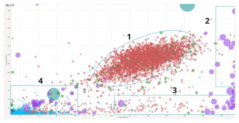
Identify cell doublets and triplets (1), cellular aggregates (2), protein aggregates (3), and plastic contaminants (4) in a cell therapeutic sample. Image Credit: Waters | Wyatt Technology
BMI: The key to sensitive, robust, early-stage particle characterization
Backgrounded membrane imaging (BMI) offers an enhanced perspective on a sample's composition, providing improved clarity, speed, and comprehensiveness. By acquiring and subtracting background images of the membrane, BMI achieves a contrast level 10 times superior to that of liquid-based techniques, such as light obscuration and flow imaging.
The Aura+ System provides comprehensive data while eliminating clogging issues and the need for in-measurement cleaning.
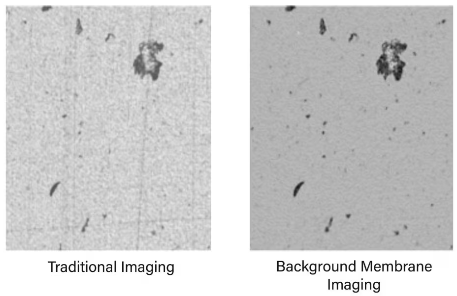
BMI achieves 10 times greater contrast than liquid-based measurements, such as light obscuration and flow imaging. Image Credit: Waters | Wyatt Technology
Get fast, definitive particle identification with three fluorescence channels
The Aura+ System is equipped with three fluorescence channels, enabling the acquisition of comprehensive data from a single experiment, which would typically necessitate three separate investigations.
Furthermore, the Aura+ System accurately distinguishes between various particle types within a given sample, facilitating more informed decision-making:
- Three fluorescence membrane microscopy (FMM) channels are available for detecting three distinct species of organic particles
- Side illumination membrane imaging (SIMI) channel is incorporated for the identification of extrinsic and inorganic particles
- It is suited for applications such as polysorbate degradation, particulate contamination, protein and antibody workflows, cell therapy workflows, AAV aggregation, AAV stability, cell therapy QC, and cell viability
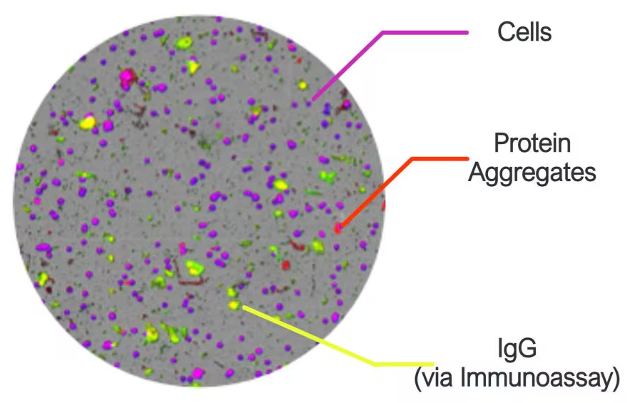
Cells, protein aggregate, and IgG (via immunoassay). Image Credit: Waters | Wyatt Technology
Obtain clear details with high magnification
The Aura+ System's high magnification capabilities enable enhanced observation of the morphological details of unlabeled particles. This facilitates clear differentiation between desired and unwanted particles.
Rapidly distinguish and count cells against Dynabeads™, assess polysorbate breakdown, and perform other analyses.
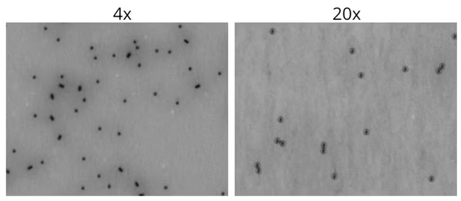
Quickly differentiate and enumerate cells versus Dynabeads™, quantify polysorbate degradation, and more. Image Credit: Waters | Wyatt Technology