The measurement of particles in cell therapy production has long presented challenges, particularly since the drug product itself is composed of particles, compounding the complexities in the process.
Waters' Aura CL System is the pioneering system that, in contrast to traditional methods, swiftly identifies and differentiates cells from protein aggregates or harmful particles, while also delivering all necessary cytometric functions. Aura CL counts, identifies, and assesses the viability of cells all in a single, simple assay.
Driven by cutting-edge technology, the Waters Aura CL System serves as the premier tool for identifying, quantifying, and characterizing subvisible particle contaminants introduced during cell therapy manufacturing. This allows users to obtain high-throughput insights earlier in the discovery phase, facilitating the development of cell therapies in a faster, more intelligent, and safer manner.
Specifications
Source: Waters | Wyatt Technology
| Specifications |
|
| Imaging Area |
24.6 mm2 |
| Optics |
4x and 10 objectives |
| Minimum volume |
5 µL (assay dependent) |
| Resolution |
1.0 pixel/µm |
| Detectable size range |
Range from >1 µm (ECD) to <5 mm (ECD) |
| BMI read time |
1 minute/sample |
| FMM read time |
30 seconds/sample |
| Software |
Particle Vue 4.x all-in-one Software suite (image capture and analysis) |
Overview
- Detect and quantify residual Dynabeads: The Aura CL System simplifies the process of locating and counting DynabeadsTM. In contrast to conventional techniques, which can be subjective, labor-intensive, inaccurate, and prone to mistakes, this advanced, fully automated instrument enables users to achieve precise counts at a rapid pace.
- ID cells and non-cells: Instead of dedicating hours to manually sifting through images or mastering intricate machine learning frameworks, the Aura CL System facilitates the straightforward identification of cells versus non-cell aggregates right from the start.
- Analyze minimal volumes: Users only require 5 µL of sample, which permits earlier characterization of particles and cells during development.
- Discover more with cutting-edge technology: By utilizing background-corrected membrane imaging (BMI) and fluorescence membrane microscopy (FMM), the Aura CL System conducts cell viability assays with clear cell identification for 96 samples of 40 μL each in under an hour.
Best used for rapid and conclusive characterization of low-volume particles in the development and manufacturing processes of cell therapy.
The first and only solution to detect and count Dynabeads
Residual Dynabeads present in a final product can undermine potency, necessitating bead analysis to adhere to stringent quality standards. Nevertheless, the application of manual hemocytometry for the detection of residual Dynabeads has proven to be inconsistent due to the difficulty in distinguishing between beads and cells.
The Waters Aura CL System employs side illumination membrane imaging (SIMI) to efficiently and accurately identify residual Dynabeads with both specificity and sensitivity, thereby reducing time and costs. This unique automated solution mitigates human errors, ensuring compliance with the highest quality standards for cell therapy lot release assays.
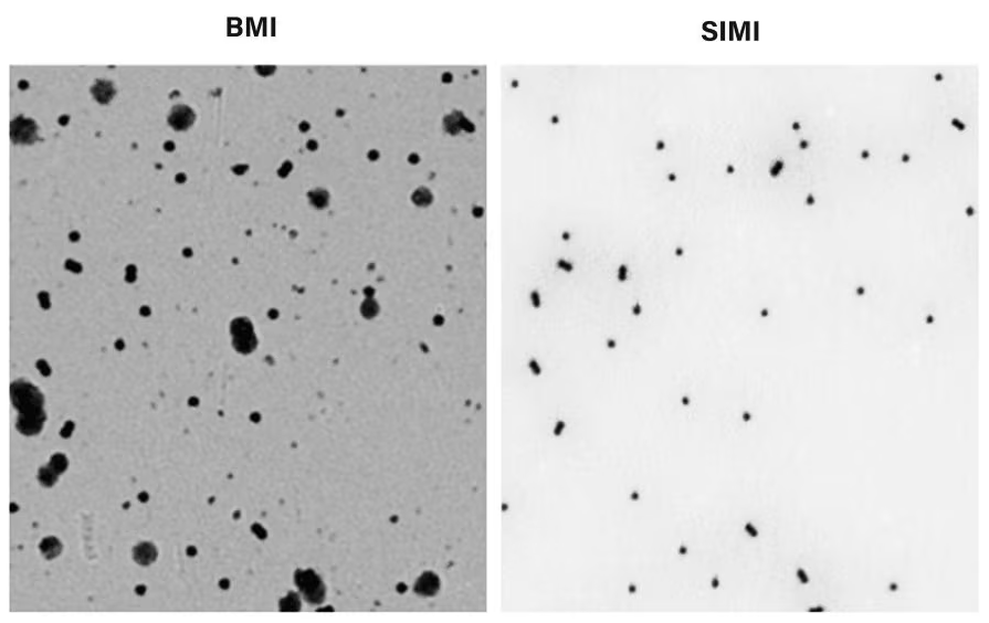
BMI reveals cells and Dynabeads, whereas SIMI reveals only Dynabeads. Image Credit: Waters | Wyatt Technology
Get high-throughput, definitive identification of viable cells
The high-throughput and accurate identification of viable cells is essential in the manufacturing of cell therapies. Nevertheless, achieving this crucial product-purity parameter has posed a challenge, as traditional methods are labor-intensive and complex - until now.
The Aura CL System eliminates uncertainty in cell viability evaluations. Users can now disregard unreliable methods, such as morphology and spectroscopy, along with intricate machine learning libraries, for assessing the sample's composition.
By labeling the particles or groups of interest with fluorescent dyes or antibodies, it becomes feasible to distinguish between cellular and non-cellular components, as well as protein and non-protein elements within complex samples.
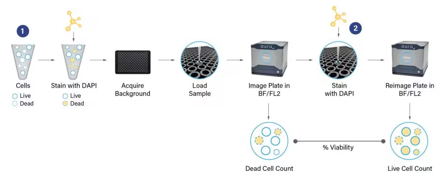
With the Aura CL System, you can achieve high-throughput and definitive identification of viable cells and take the guesswork out of cell viability assessments. Image Credit: Waters | Wyatt Technology