Introduction to hypoxia
Symptoms, types and clinical presentation of hypoxia
Etiopathogenesis of hypoxia
Hypoxia diagnosis
Hypoxia management
References
Further reading
Introduction to hypoxia
In medicine, hypoxia is a condition in which the human body tissues are not oxygenated sufficiently to maintain adequate homeostasis, resulting from inadequate oxygen delivery to the tissues due to either low blood supply or low oxygen content in the blood. Oxygen deprivation can severely affect various body cells that perform essential biological processes. The term anoxia is used in the case of complete deprivation of oxygen supply in the body.
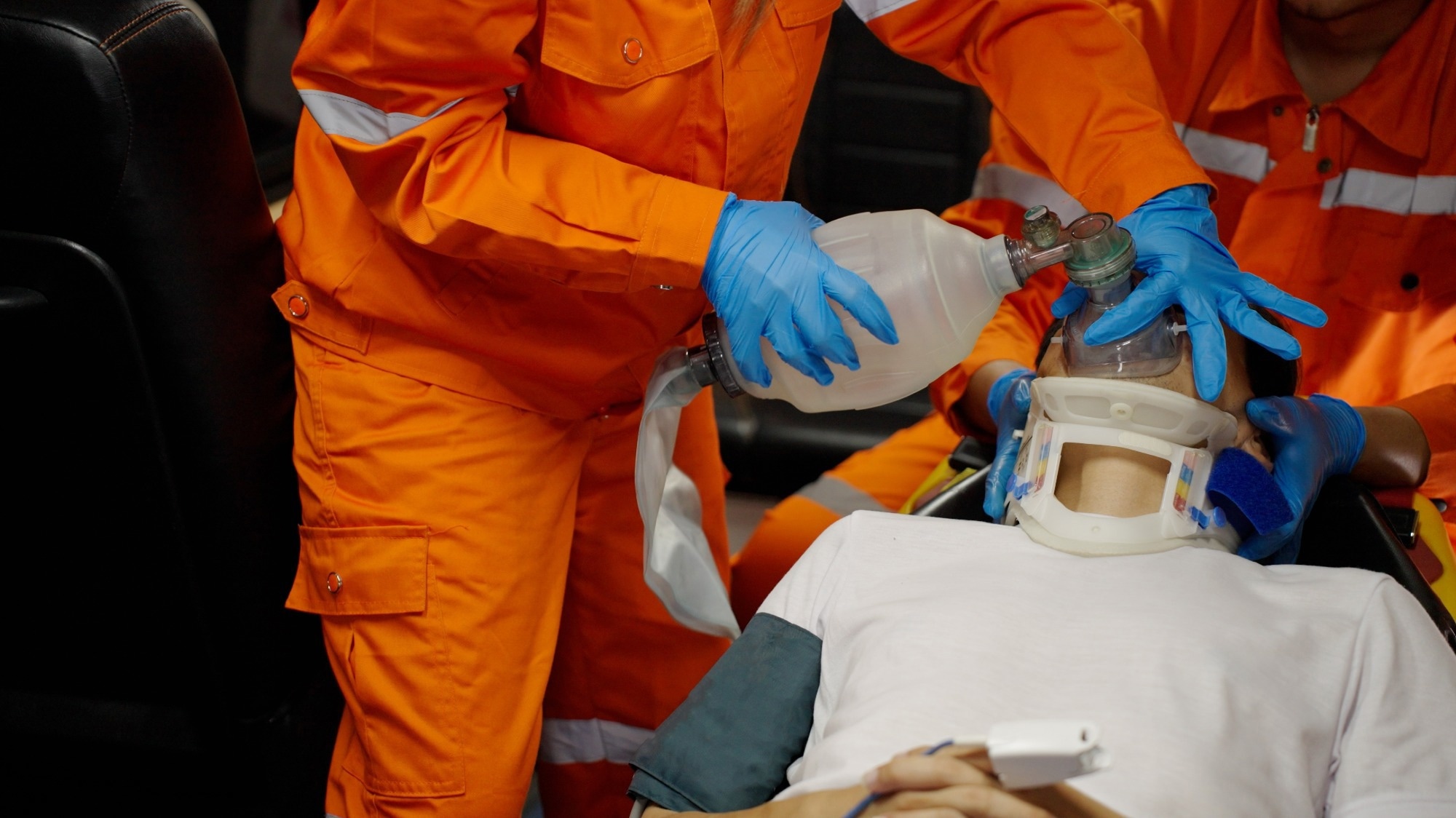 Image Credit: comzeal images / Shutterstock
Symptoms, types and clinical presentation of hypoxia
Hypoxia can vary in intensity, presenting in mild to severe forms and acute and chronic forms, and can be classified as local if a specific area of the body is affected and generalized if the entire body is affected. In chronic hypoxia, an individual may present with dyspnea on exertion, whereas, in acute hypoxia, both dyspnea and tachypnea are observed.
In some cases, tachycardia, increased myocardial contractility, and cardiac output are observed as compensatory mechanisms adopted by the body to increase the output of circulating oxygenated blood to the vital organs by decreasing the amount of blood supplied to the peripheral tissues, leading to cyanosis in peripheral areas. In addition, the oxygen saturation may be lower in hypoxia.
In moderate hypoxia, neurologic manifestations, including restlessness, headache, and confusion, are observed. Severe hypoxia can result in cyanosis and tachycardia, altered mentation and coma, and even death if not corrected quickly. Stridor (noisy breathing) can be heard in cases of upper airway obstruction. Productive cough and fever are observed in cases of lung infection, leg edema, and orthopnea in cases of heart failure, and chest pain and unilateral leg swelling may point to pulmonary embolism as a cause of hypoxia.
Symptoms of mild cerebral hypoxia (decreased oxygen supply to the brain) include inattentiveness, poor judgment, memory loss, and decreased motor coordination since brain cells are extremely sensitive to oxygen deprivation and can begin to die within five minutes after the oxygen supply has been cut off. Prolonged hypoxia can also result in seizures.
Intrauterine hypoxia is a significant clinical challenge in obstetrics observed among pregnant women residing at high altitudes and/or with cardiovascular diseases. Altered placental development and spiral artery remodeling leading to placental insufficiency and dysfunction are other causes. Both conditions can impact normal maternal cardiovascular homeostasis leading to preeclampsia and/or impairing the transfer of oxygen supply to the fetus resulting in fetal growth restrictions.
Lung auscultation can yield helpful information to diagnose the underlying condition that presents clinically with hypoxia. Bilateral basilar crackles may indicate pulmonary edema or volume overload, other signs of which include jugular venous distention and lower limb edema. Wheezing and rhonchi can be found in obstructive lung disease, and absent unilateral air entry can be caused by either massive pleural effusion or pneumothorax.
Further, chest percussion can help differentiate between pleural effusion and pneumothorax by revealing dullness in cases of pleural effusion and hyper-resonance in cases of pneumothorax. Clear lung fields in a setting of hypoxia should raise suspicion of pulmonary embolism, especially if the patient is tachycardic and has evidence of deep vein thrombosis (DVT).
Etiopathogenesis of hypoxia
Hypoxia could result from anemia, in which the amount of functional hemoglobin is decreased, affecting the oxygen-carrying ability of the blood. Another cause is carbon monoxide poisoning, wherein the chemical bonds to oxygen receptors on red blood cells are affected, resulting in cerebral hypoxia. In addition, drowning, strangling, choking, suffocation, cardiac arrest, head trauma, and general anesthesia complications can lead to cerebral hypoxia.
Hypoxia may also be caused by conditions such as asthma, bronchitis, chronic obstructive pulmonary disease (COPD), emphysema, pneumonia (bacterial, viral), congestive heart failure, myocardial infarction, pneumothorax, pulmonary edema, pulmonary embolism, pulmonary hypertension, pulmonary fibrosis, and sleep apnea.
Further, healthy people may also suffer from hypoxia, e.g., due to travel to high altitudes where the partial pressure of oxygen in the inhaled air and oxygen tension is low, and during deep sea diving in case the breathing gases have been incorrectly prepared or if rusty cylinders in their gas tanks have extracted oxygen.
To have oxygen carried by hemoglobin, direct interaction between red blood cells (RBCs) in pulmonary capillaries and the air in the alveoli is required, which can be compromised at either of the following points: (i) during perfusion or blood flow to the lungs, (ii) during ventilation or airflow to the alveoli, and (iii) during diffusion or gaseous exchange via interstitial tissues.
Hypoventilation may be caused due to several reasons, including (i) airway obstruction, either proximal as in laryngeal edema or foreign body inhalation or distal as in bronchial asthma or COPD, (ii) impaired respiratory drive as in cases of deep sedation or coma, (iii) restricted movement of the chest wall as in obesity hypoventilation syndrome, circumferential burns, massive ascites, or ankylosing spondylitis, and (iv) neuromuscular diseases, such as myasthenia gravis, muscular dystrophy, amyotrophic lateral sclerosis, or phrenic nerve injuries.
A lower ventilation-perfusion (V/Q) ratio is observed due to impaired ventilation or high perfusion in chronic bronchitis and airway obstruction by mucus plugs and pulmonary that impair ventilation. Increased V/Q ratio is observed due to impaired perfusion in pulmonary embolism or increased ventilation in emphysema, wherein the surface area available for gas exchange is decreased.
Right to left shunt or crossing of blood from the right to the left side of the heart without being oxygenated occurs as anatomic shunts or physiologic shunts. Anatomic shunts are observed in cases wherein blood bypasses the alveoli, e.g., intracardiac shunts, pulmonary arteriovenous malformations, fistulas, and hepato-pulmonary syndrome. Physiologic shunting is observed when blood passes through non-ventilated alveoli, as in pneumonia, atelectasis, and acute respiratory distress syndrome (ARDS). In addition, oxygen diffusion is impaired between the alveolus and the pulmonary capillaries in cases of interstitial edema, interstitial inflammation, or fibrosis, as in pulmonary edema and interstitial lung disease.
Hypoxia diagnosis
Pulse oximetry is performed to evaluate arterial oxygen saturation (SaO2), i.e., the amount of oxygen bound to hemoglobin in arterial blood and resting SaO2 ≤95% or exercise desaturation ≥ five percent is considered abnormal. In addition, arterial blood gas evaluation is a valuable tool for evaluating hypoxemia and can yield additional information, such as the partial pressure of CO2 (PCO2), which can shed light on the etiology of hypoxia. PCO2 is elevated in hypoventilation cases and cases of acute hypoxia secondary to tachypnea and washout of CO2.
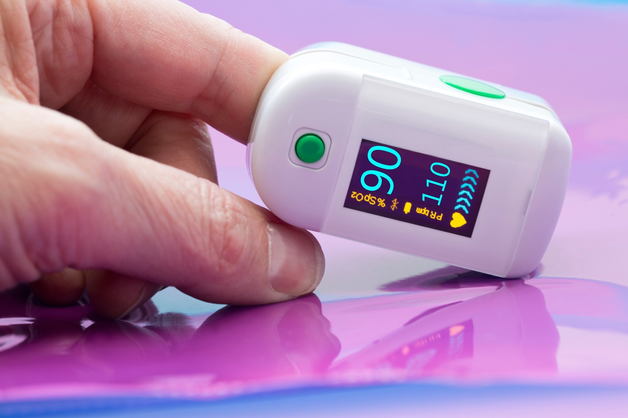 Portable finger pulse oximeter. Image Credit: Beate Panosch / Shutterstock
Imaging studies of the chest, such as chest x-rays or computed tomography (CT), help in identifying the cause of hypoxia, e.g., pneumonia, pulmonary edema, hyperinflated lungs in COPD, and other conditions. CT chest can yield detailed images that outline the exact pathology, and CT angiograms of the chest are particularly important in detecting pulmonary embolism.
Another modality is the VQ scan to detect ventilation-perfusion mismatch in acute or chronic pulmonary embolism. The VQ scan can be particularly useful when renal failure or allergy to iodinated contrast increases the risks of CT angiography. Pulmonary function tests (PFT), nocturnal trend oximetry, six-minute walk test, and hemoglobin concentration tests are performed to assess chronic hypoxia.
Hypoxia management
Management of hypoxia can be broadly classified under three categories which are: (i) maintaining patent airways, (ii) increasing the oxygen content of the inspired air, and (iii) improving the diffusion capacity. The patency of upper airways can be ensured with good suctioning, maneuvers that prevent occlusion of the throat (head tilt and jaw thrust), and endotracheal tube placement or tracheostomy when necessary.
In chronic conditions such as obesity hyperventilation syndrome, maintaining patent airways can be achieved with positive pressure ventilation like CPAP (continuous positive airway pressure) and BiPAP (bilevel positive airway pressure). Bronchodilators and aggressive pulmonary hygiene, such as chest physiotherapy, the flutter valve, and incentive spirometry, can be used to maintain the patency of the lower airways.
The fraction of inspired oxygen must be increased in cases of PaO2 <60 or SaO2 <90 by using low-flow devices such as nasal cannulas, reservoir cannulas (oxymizers), partial-rebreather masks, non-rebreather masks, and simple face masks. High-flow devices such as venturi masks, high-flow nasal cannulas, and air/oxygen blenders could also be used.
The underlying cause of respiratory failure must be identified and treated. Diuretics can be used in cases of pulmonary edema and steroids in some instances of interstitial lung disease. Extracorporeal membrane oxygenation (ECMO) can be used as an ultimate method of increasing oxygenation. Smoking cessation and quitting tobacco can help increase lung function and prevent further damage to your lungs.
References
- www.faa.gov/pilots/safety/pilotsafetybrochures/media/hypoxia.pdf - HYPOXIA
- http://link.springer.com/chapter/10.1007%2F978-0-387-75246-4_97 - Hypoxemia and Hypoxia, Common Surgical Diseases pp 391–394
- https://www.ncbi.nlm.nih.gov/books/NBK482316/ - Hypoxia
- https://my.clevelandclinic.org/health/diseases/23063-hypoxia - Hypoxia
- https://www.dovepress.com/intrauterine-hypoxia-clinical-consequences-and-therapeutic-perspective-peer-reviewed-fulltext-article-RRN - Intrauterine hypoxia: clinical consequences and therapeutic perspectives
Further Reading
Last Updated: Sep 6, 2022
 Can smoking while pregnant harm your child’s teeth? New research suggests yes