Introduction
How SPR Supports Drug Discovery?
The Role of Molecular Docking in Understanding Binding Mechanisms
Combining SPR and Molecular Docking: Insights From Tryptase Inhibitor Research
Implications For Drug Discovery and Future Applications
References and Further Reading
Research on tryptase inhibitors demonstrates how combining experimental binding measurements with computational predictions reveals differences in drug–target affinity, binding stoichiometry, and molecular recognition. These findings highlight the value of integrating biophysical and structural evidence to guide inhibitor development and lead optimization.
 Image credit: S. Singha/Shutterstock.com
Introduction
Identifying interactions between small molecules and biological targets is critical during a drug discovery process. Determining a small molecule's binding affinity to a biological target is important for assessing its drug potential. Likewise, knowing the rate at which binding occurs and how long the small molecule is bound to the biological target is critical in the drug discovery process.
Binding affinity defines how strongly a small molecule binds to a biological target, whereas binding kinetics defines the rate at which small molecules associate with and dissociate from a biological target. Surface plasmon resonance (SPR) and molecular docking are two complementary methods for characterizing these interactions. SPR is an experimental method used to determine binding affinity and kinetics. On the other hand, molecular docking is a computational method that predicts possible interactions between small molecules and biological targets.1,4
Download your PDF copy now!
How SPR Supports Drug Discovery?
SPR is an optical method that detects real-time changes in the refractive index near the sensor surface when molecules bind at or near it. In a standard SPR experiment, one interaction partner, termed the ligand, is immobilized or captured on the sensor chip while the other participant, known as the analyte, passes over the surface of the sensor. Binding changes the sensor surface's optical properties and generates a measurable signal without any need for a fluorescent or radioactive label.1,4
The most important benefit of SPR lies in its ability to calculate key binding parameters of the complex. Specifically, the association rate constant (kon) indicates how fast two molecules bind together, while the dissociation rate constant (koff) states the rate of separation of the formed complex. The equilibrium dissociation constant (KD) represents binding affinity and, for a simple 1:1 interaction, is calculated by KD = koff/kon. Lower KD means stronger binding in general, while the dissociation rate provides information about the stability of the molecular complex and its residence time, which is the duration a compound remains associated with its target.1,4
SPR measurements can also help scientists understand binding stoichiometry, which refers to the number of ligand molecules bound to a target protein or protein complex. However, valid interpretation is contingent upon appropriate experimental conditions such as ligand immobilization, analyte concentration, flow rate, and the properties of the sensor surface. Mass transport limitations, which occur when analyte delivery to the sensor surface restricts binding, can distort kinetic measurements. Therefore, appropriate assay design and mathematical modeling are necessary to obtain reproducible interaction parameters.1
Multiparametric SPR profiling can further support small-molecule development by examining affinity, association and dissociation kinetics, and selectivity across different targets. Bruker's SPR resources include an application report on bacterial glutaminyl cyclase inhibitors that illustrates how interaction profiling can contribute to compound characterization and lead optimization.2
Surface Plasmon Resonance (SPR) Technology, Explained
Video credit: @SartoriusGlobal/youtube.com
The Role of Molecular Docking in Understanding Binding Mechanisms
Molecular docking is a computational approach used to predict how small molecules interact with specific binding regions of target proteins. The method evaluates possible ligand orientations within a protein-binding pocket and estimates their compatibility using geometric and energetic scoring functions.1
Docking can provide structural explanations for differences in experimentally observed binding affinities. However, docking predictions do not independently establish whether a compound binds to its intended target. Scoring functions provide estimates rather than direct experimental measurements, and their accuracy can be influenced by protein flexibility, solvent effects, protonation states, and the treatment of molecular interactions. Predicted binding poses should therefore be interpreted as hypotheses requiring experimental validation. Combining docking with biophysical measurements and structural techniques can strengthen the interpretation of molecular recognition mechanisms.1,4
Combining SPR and Molecular Docking: Insights From Tryptase Inhibitor Research
According to a 2025 study by Porta et al., SPR, molecular docking, and X-ray crystallography can work together to assess tryptase inhibitors. The authors analyzed three different compounds previously mentioned in the literature: compound #1, a monovalent inhibitor; compound #2m, another monovalent inhibitor; and compound #2d, a bivalent inhibitor. The investigation focused on their binding affinity, interaction kinetics, and binding arrangements within human tryptase, a tetrameric serine protease containing four binding pockets.1
The researchers initially used SPR to experimentally characterize the interactions between the inhibitors and immobilized human tryptase. All three compounds demonstrated concentration-dependent binding. The measured KD revealed substantial differences in affinity. Bivalent compound #2d demonstrated the strongest binding, with a mean KD of 2.1 nM, followed by compound #1 at 12.3 nM and compound #2m at 81 nM.1
The kinetic measurements provided additional insight into these differences. Compound #1 demonstrated a faster association rate than compound #2m, contributing to its stronger affinity despite a faster dissociation rate. Similarly, the higher affinity of bivalent compound #2d compared with its corresponding monovalent compound was primarily associated with a 38-fold faster association rate, while their dissociation rates were relatively similar. These findings indicated that differences in binding affinity were influenced by association kinetics rather than simply by the duration of molecular interactions.1
SPR measurements also provided information about binding stoichiometry. The researchers observed comparable maximum binding responses (Rmax) after accounting for differences in molecular weight. These findings suggested that all four binding pockets of the tryptase tetramer were accessible to the inhibitors. Monovalent compounds #1 and #2m were consistent with a 4:1 stoichiometry, with four inhibitor molecules binding to one enzyme tetramer. In contrast, bivalent compound #2d was consistent with a 2:1 stoichiometry, indicating that two inhibitor molecules could occupy the four binding pockets by simultaneously engaging adjacent sites.1
Molecular docking was then used to clarify the molecular basis of the experimental results. The simulation results indicated certain variations in the binding patterns for the studied compounds. In particular, compound #1 was predicted to bind through five hydrogen bonds involving residues such as Ser209, His59, Lys66, Gly207, and Ser204. Compound #2m was predicted to possess fewer hydrogen bonds as well as other polar interactions, which may contribute to the lower binding affinity for this compound. Compound #2d was predicted to use two adjacent sites for binding.1
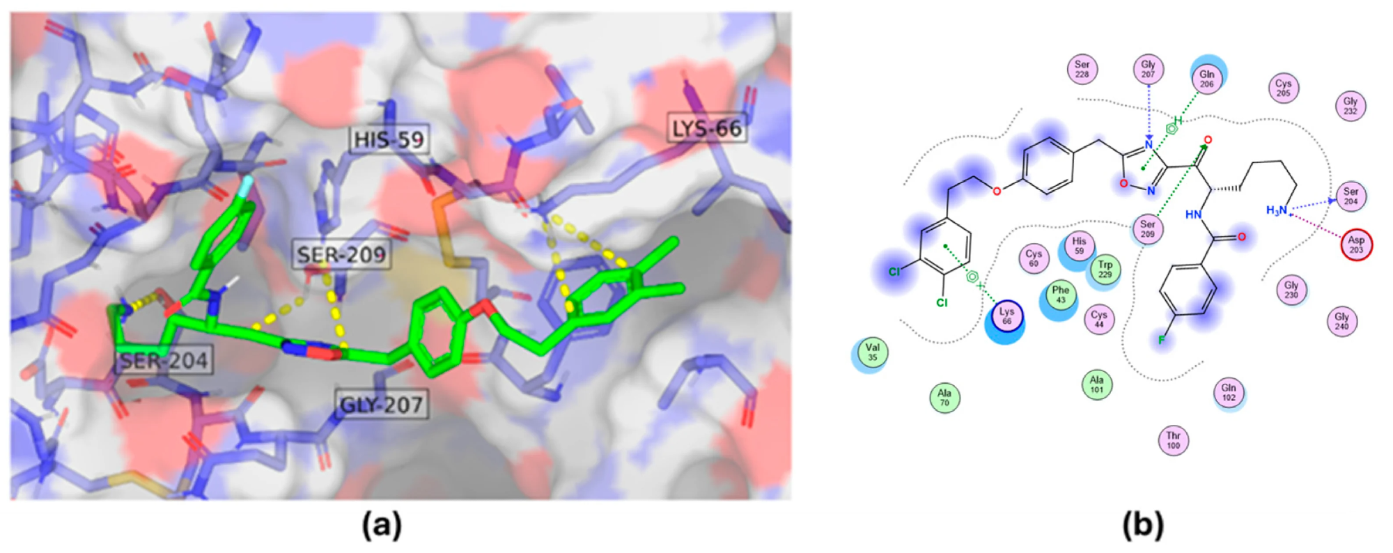 Molecular interactions of a tryptase inhibitor.** Three-dimensional binding model (a) and two-dimensional interaction diagram (b) illustrating the inhibitor's predicted binding orientation and interactions with key amino acid residues in the tryptase binding pocket. Image credit: Porta et al. (2025).
The obtained docking results helped explain the observed binding stoichiometry. Monovalent compounds were predicted to occupy individual binding sites separately, while the bivalent compound would bind using two adjacent binding sites within the same tetramer. These predictions explain the determined 4:1 and 2:1 binding ratios based on the experimental results.1
To provide further structural validation, the researchers determined X-ray crystal structures of human tryptase beta-2 (hTPSB2) in its unbound form and in complex with compound #1. The complex structure was resolved at 2.06 Å, allowing the investigators to examine the inhibitor's binding orientation and molecular contacts. The experimentally determined structure confirmed the binding mode and key interactions predicted through docking. The study also compared these findings with previously reported structural information for bivalent compound #2d.1
Implications For Drug Discovery and Future Applications
Integrating SPR and molecular docking can improve compound characterization by connecting quantitative binding measurements with predicted molecular interactions. SPR identifies differences in affinity, association rates, and dissociation rates, while docking helps investigate the structural features potentially responsible for these differences. This combination can support the selection of promising compounds, guide chemical modifications, and help researchers investigate binding stoichiometry and molecular recognition during early drug development.1,2,4
Additional biophysical information, particularly binding thermodynamics, may further strengthen this approach. Thermodynamic profiling examines the energetic contributions associated with molecular complex formation, including enthalpy and entropy. These parameters can provide insights into the forces driving ligand binding and help distinguish compounds with comparable affinities but different interaction profiles. Bruker's 2025 webinar on multiplexed SPR-based thermodynamic profiling illustrates the potential for evaluating affinity, selectivity, and thermodynamic characteristics across multiple compounds and protein targets within an experimental workflow.3
Watch the full webinar from Bruker Dynamic Biosensors: DISCOVER the fast track to thermodynamics: profiling with SPR
Notably, several limitations must be considered. Docking predictions depend on computational assumptions and may not fully represent protein flexibility, solvent conditions, or changes occurring during molecular binding. SPR measurements can also be affected by immobilization procedures, nonspecific interactions, mass transport limitations, and assay conditions. Functional measurements should be performed to evaluate compound potential; this includes selectivity assays, cellular experiments, and preclinical research. Their integration offers a useful framework for investigating binding mechanisms and making evidence-based decisions during drug discovery and lead optimization.1,4
References and Further Reading
- Porta, A., Manelfi, C., Talarico, C., Beccari, A. R., Brindisi, M., Summa, V., Iaconis, D., Gobbi, M., & Beeg, M. (2025). Integrating Surface Plasmon Resonance and Docking Analysis for Mechanistic Insights of Tryptase Inhibitors. Molecules. 30(6), 1338. DOI: 10.3390/molecules30061338, https://www.mdpi.com/1420-3049/30/6/1338
- Bruker. Surface plasmon resonance (SPR). https://www.bruker.com/en/products-and-solutions/biosensors-for-molecular-interactions/surface-plasmon-resonance
- Bruker. Discover the fast track to thermodynamics: Profiling with SPR. https://www.bruker.com/de/news-and-events/webinars/2025/discover-the-fast-track-to-thermodynamics-profiling-with-spr
- Singh, D. K., Tiwari, S., & Kripalata, K. (2026). Surface plasmon resonance in drug discovery: Advances in protein-protein interaction and its kinetic analysis. Discover Chemistry. 3. Article 337. DOI: 10.1007/s44371-026-00797-0, https://link.springer.com/article/10.1007/s44371-026-00797-0
Last Updated: Nov 10, 2026