Förster resonance energy transfer (FRET) is a technique that depends on the distance between two flourophores and the transfer of energy from one excited fluorophore to another by intermolecular long-scale dipole–dipole coupling. FRET can be measured in numerous ways. One of the ways of quantitating FRET between two similar fluorophores is called homo FRET.
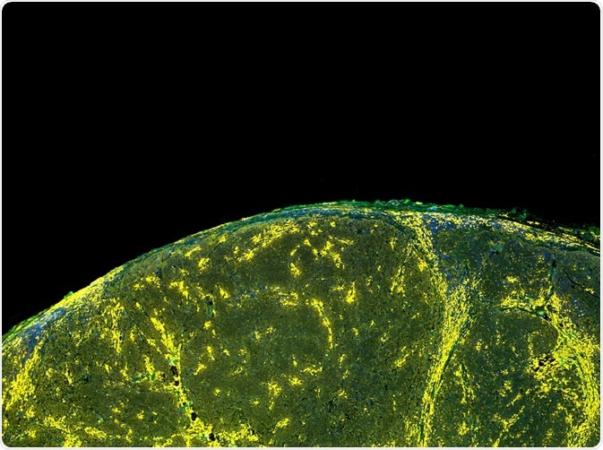
Credit: Carl Dupont/ Shutterstock.com
Homo FRET is a non-radiative transfer between two similar probes that display an intersection of emission and absorption spectra. Therefore, it is utilized to quantify interactions between proteins with one fluorescent probe. Homo FRET is advantageous over hetero FRET as it does not cause any modifications in the lifetime of the donor fluorophore. It transfers the excited energy from the donor to the same kind of acceptor fluorophore. This technique has set a reliable beginning for future speculations to solve problems in life science.
Study of molecular self-assembly
Organization of proteins and lipids in cell membranes is an important attribute of various biological behavior and dysfunctions such as fundamental cell signaling and protein aggregation. For analysis of the process of self-assembly in live cells, homo FRET approach is used to detect and measure the molecular clusters in the cells through fluorescence anisotropy imaging microscopy. Therefore, it provides a strong understanding of the molecular functioning of live and diseased cells.
Homo FRET imaging is applied to detect the self-assembly of glycosylphosphatidylinositol (GPI)-anchored proteins in live cellular membranes. The depolarization induced by homo FRET is higher in arbitrarily distributed proteins than in domain-bound proteins. Experiments on membranes and lipids arrangement reveal that there is uniform anisotropic distribution in cells expressing fluorescent-labeled GPI-anchored proteins. A decrease in fluorophore density due to photobleaching results in an increase in anisotropy in these cells.
Homo FRET for examining insulin package
A cell-based assay involving fluorescent proteins for homo FRET detection is used to examine insulin granule packaging in live cells. Resistance to insulin or less insulin production due to disturbance in metabolism leads to diabetes mellitus. As diabetes is a common health disorder prevalent in the world population, there is a constant need to find new therapeutic approaches to curb the disease.
It is analyzed that a homo FRET and fluorescence polarization approach can monitor the level of packaging developed insulin into a compact core granule. The method consists of a red fluorophore (mCherry) combined with the precursor of insulin and is made to express in the cells. The cells are treated with bafilomycin to block formation of insulin granules. The approach shows that high concentration of bafilomycin leads to fewer granularities of insulin and vice versa. Ultimately, homo FRET technique established a new cell-based biosensor to detect compounds that alter insulin granule packaging.
Determination of protein cluster sizes
Homo FRET based on fluorescence anisotropy methods is useful in the study of identical protein clusters in live cells. To determine the cluster size and distribution of similar proteins, cells expressing green fluorescent protein (GFP) are analyzed. However, analysis of homo-FRET signals is difficult due to the fact that both the mutual introduction of the fluorophores and the total count of fluorophores present in every cluster tend to affect the fluorescence anisotropy in the same manner.
Studies of GPI-anchor were conducted to investigate the clustering and affinity properties by using homo FRET approach. Till date, the size of cluster has been determined using conventional approaches (microspectroscopic techniques) by deriving from a plot of anisotropy versus photobleaching level. But the homo FRET-based fluorescent anisotropic method allows to measure heterogeneities in protein clustering within the cellular components.
A GPI-anchor is fused with GFP and expressed in many distinct locations in the cell, and most prominently in plasma membrane and Golgi apparatus. The early anisotropic decay is rapid, which indicates a very high efficiency in energy transfer and demonstrating that the distance between the fluorophores is insignificant, allowing interaction between the GFPs.
Therefore, it is understood that GPI–GFP’s clustering behavior depends on cellular localizations and is mostly found in plasma membrane consisting of small clusters of size longer than dimers, whereas in Golgi, GPI–GFP is found as monomers.
Biosensors in imaging signaling events
Biosensors with homo FRET multiplexed imaging technique is a powerful and successful approach to observe the spatiotemporal association of signaling pathways in a living cell. It is demonstrated that the homo-FRET between the mCherry-labeled pleckstrin homology domains of Akt can be used to observe the accumulation of 3'-phosphoinositide in living cells.
The phosphorylated phosphatidylinositol results in phosphoinositides that are lipids bound to membrane play an essential role in signaling the numerous cellular processes such as membrane trafficking, chemotaxis, and cytoskeletal regulation. This technique provides a useful approach to reveal the connection between these signaling pathways in facilitating chemotaxis. Also, to exhibit the global analysis of fluorescence anisotropy measurements can be utilized to compute and measure the accumulation of 3'-phosphoinositide at the membranes.
Further Reading
Last Updated: May 24, 2019