Introduction
Structure and Composition
Formation and Maintenance
Biological Functions
The Endothelial Glycocalyx
Role in Health and Disease
Methods to Study the Glycocalyx
Future Outlook
References
The glycocalyx emerges as an active endothelial system whose structure, metabolism, and shear-dependent regulation integrate mechanical forces with vascular signaling. Disruption of this system provides a unifying mechanism linking endothelial dysfunction to inflammation, barrier failure, and cardiometabolic disease.
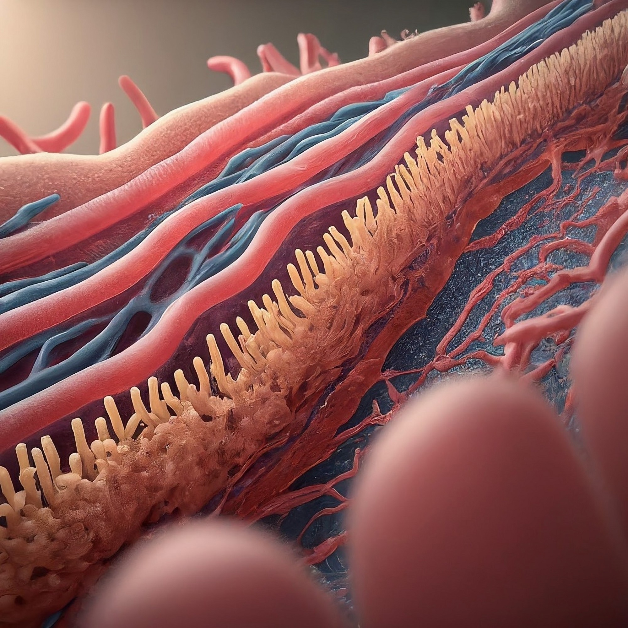 Image credit: Shutterstock AI/Shutterstock.com
For decades, researchers conceptualized the vascular endothelium as a simple, non-thrombogenic layer lining the blood vessels, a passive barrier separating the circulating blood from the vessel wall. More recent research has debunked this reductionist view by elucidating the endothelial glycocalyx (eGCX), a complex, gel-like "glycoscape" that coats the luminal surface of endothelial cells. 1
It is important to clarify that the term “GlycoLax” is a common pharmaceutical trade name for polyethylene glycol–based laxatives and is unrelated to vascular biology; the correct biological term discussed here is the endothelial glycocalyx. 2
Studies have shown that the glycocalyx is a dynamically regulated organelle that alters vascular homeostasis. It is now known to serve as the primary mechanotransducer of hemodynamic shear stress, a gatekeeper of vascular permeability, and a crucial modulator of the immune response.
This article synthesizes recent research to present a comprehensive overview of the glycocalyx, detailing its molecular architecture and non-template-driven biosynthesis and highlighting how these processes connect vascular health to cellular metabolism.
Furthermore, it unravels how the pathological "shedding" of this layer drives sepsis, diabetes, and atherosclerosis, and evaluates emerging therapeutic strategies, from sulodexide to synthetic glycoscapes, that aim to restore this fragile yet vital barrier.
Introduction
The endothelial glycocalyx is a multicomponent, carbohydrate-rich layer that coats the luminal surface of endothelial cells. Formerly considered an artifact of microscopy, it is now identified as a critical regulator of the cell's interaction with its environment.1
A growing body of literature suggests that the glycocalyx functions as a physical barrier and a transduction interface, mediating the exchange of information and force between circulating blood and the vessel wall. Its integrity is the defining feature of a healthy vascular interface. At the same time, its degradation is increasingly recognized as a trigger for diverse pathologies ranging from acute inflammation to chronic vascular disease.1
Structure and Composition
Physically, the glycocalyx resembles a "bush-like" meshwork composed of a proteoglycan backbone and a canopy of glycosaminoglycans (GAGs). The layer's structural foundation is provided by transmembrane proteoglycans, primarily syndecans (particularly Syndecan-1), which anchor the layer to the cytoskeleton, and GPI-anchored glypicans (e.g., Glypican-1), which localize to lipid rafts.1,2
Extending from these proteoglycan cores are long, negatively charged GAG chains, predominantly heparan sulfate (50–90 %) and chondroitin sulfate, which create a vast reservoir for binding plasma ligands.1
Interwoven within this mesh is hyaluronan (hyaluronic acid), an extremely long, non-sulfated polymer that does not bind covalently to core proteins but instead interacts with surface receptors (e.g., CD44) to provide hydration and structural volume.1,2
Formation and Maintenance
Recent research highlights that, unlike most structural proteins (which are encoded in the host's DNA), the glycocalyx is synthesized through a complex, non-template-driven metabolic process.3
The glycocalyx's proteoglycan cores are synthesized in the Endoplasmic Reticulum (ER), while the GAG chains are elongated and modified by specific enzymes (such as EXT1/2 and sulfotransferases) in the Golgi apparatus.3 Hyaluronan has recently been shown to demonstrate a notable exception to this synthesis pathway; it is synthesized directly at the plasma membrane by Hyaluronan Synthases (HAS), with HAS2 being the dominant endothelial isoform. 4
Studies have shown that hemodynamic forces dynamically regulate the biological maintenance of the glycocalyx. Specifically, laminar shear stress activates the transcription factor KLF2, which upregulates HAS2 expression and redistributes endothelial glucose flux from glycolysis toward UDP-sugar biosynthesis, thereby supplying substrates required for glycocalyx regeneration. 9
Biological Functions
Mechanistic investigations have identified three main glycocalyx functions: 1. Protection, 2. Permeability modulation, and 3. Mechanotransduction (conversion of physical environmental cues into biochemical signals).1,3,5
- The layer's dense, negatively charged meshwork has been observed to repel red blood cells and mask shorter adhesion molecules (like ICAM-1), thereby preventing inadvertent leukocyte attachment.5
- The layer's structural configuration further acts as a molecular sieve, restricting the extravasation of fluids and macromolecules like albumin.3,5
- The most recently described function of the glycocalyx is that of mechanotransduction. Research suggests that the drag force of blood flow bends heparan sulfate chains, transmitting tension through syndecan core proteins to the actin cytoskeleton, which stimulates endothelial nitric oxide synthase (eNOS) activity and nitric oxide (NO) release.8
The Endothelial Glycocalyx
In the vascular system, the endothelial glycocalyx (eGCX) is the primary determinant of vascular health. A growing body of evidence indicates that the eGCX is not uniform across the body, with significant thickness and compositional heterogeneity among organs.1,5
In the renal glomerulus, the highly anionic glycocalyx contributes substantially to charge-selective filtration, and its degradation has been directly linked to albuminuria in both experimental and clinical studies. 3
In contrast, in the blood-brain barrier (BBB), a dense glycocalyx has been observed to work in concert with tight junctions to strictly limit the entry of potentially neurotoxic elements. Epidemiological and experimental studies now link BBB-associated eGCX degradation to neuroinflammation and cerebral edema.1,6
Role in Health and Disease
The degradation or "shedding" of the glycocalyx is now established as a hallmark of systemic disease. In sepsis, inflammatory cytokines and oxidative stress activate enzymes such as heparanase and matrix metalloproteinases (MMPs), leading to the release of syndecan-1 and heparan sulfate fragments into the circulation.2,6
A 2025 meta-analysis confirmed that elevated plasma levels of Syndecan-1 are significantly associated with increased mortality in sepsis patients (Odds Ratio = 2.04), validating glycocalyx degradation as a prognostic indicator of endothelial injury.7
Furthermore, in diabetes and atherosclerosis, disturbed shear stress and chronic metabolic dysfunction impair glycocalyx biosynthesis, exposing the endothelium to lipoprotein infiltration and leukocyte adhesion, thereby accelerating plaque formation.6,9
Methods to Study the Glycocalyx
Traditional electron microscopy (EM) often dehydrates samples, causing glycocalyx collapse and underestimation of thickness. To overcome this, cryo-electron microscopy and Sidestream Dark Field (SDF) imaging are now used to preserve the hydrated endothelial surface layer in vivo.8
Recently, super-resolution techniques such as Resolution Enhancement by Sequential Imaging (RESI) combined with metabolic labeling have achieved ångström-level resolution, enabling visualization of individual sugar residues within intact cell-surface glycans.2
Future Outlook
While earlier work emphasized visualization, the field is now shifting toward intervention. Glycocalyx-targeted therapeutics such as sulodexide, a mixture of heparan and dermatan sulfates, have demonstrated efficacy in restoring glycocalyx integrity, reducing endothelial permeability, and improving outcomes in experimental and clinical sepsis.1
Bioengineers are also developing synthetic glycoscapes, engineered nanomaterials designed to reconstitute endothelial surface layers, representing a promising frontier for acute vascular protection and precision therapeutics.2
References
- Jin, J., et al. (2021). The Structure and Function of the Glycocalyx and Its Connection With Blood-Brain Barrier. Frontiers in Cellular Neuroscience, 15. DOI:10.3389/fncel.2021.739699, https://www.frontiersin.org/journals/cellular-neuroscience/articles/10.3389/fncel.2021.739699/full
- Machin, D. R., Sabouri, M., Zheng, X., & Donato, A. J. (2023). Therapeutic strategies targeting the endothelial glycocalyx. Current Opinion in Clinical Nutrition & Metabolic Care, 26(6), 543–550. DOI:10.1097/MCO.0000000000000973, https://journals.lww.com/co-clinicalnutrition/Abstract/2023/11000/Therapeutic_strategies_targeting_the.12.aspx
- Wang, G., et al. (2020). Shear Stress Regulation of Endothelial Glycocalyx Structure Is Determined by Glucobiosynthesis. Arteriosclerosis, Thrombosis, and Vascular Biology, 40(2), 350–364. DOI:10.1161/ATVBAHA.119.313399, https://www.ahajournals.org/doi/10.1161/ATVBAHA.119.313399
- Milusev, A., Rieben, R., & Sorvillo, N. (2022). The Endothelial Glycocalyx: A Possible Therapeutic Target in Cardiovascular Disorders. Frontiers in Cardiovascular Medicine, 9. DOI:10.3389/fcvm.2022.897087, https://www.frontiersin.org/journals/cardiovascular-medicine/articles/10.3389/fcvm.2022.897087/full
- Tarbell, J. M., & Pahakis, M. Y. (2006). Mechanotransduction and the glycocalyx. Journal of Internal Medicine, 259(4), 339–350. DOI:10.1111/j.1365-2796.2006.01620.x, https://onlinelibrary.wiley.com/doi/10.1111/j.1365-2796.2006.01620.x
- Daniyarova, K. R., et al. (2025). Glycocalyx and Endothelial Biomarkers as Prognostic Indicators in Sepsis: A Systematic Review and Meta‐Analysis. MicrobiologyOpen, 14(6). DOI:10.1002/mbo3.70155, https://onlinelibrary.wiley.com/doi/10.1002/mbo3.70155
- Ying, J., et al. (2023). Sulodexide improves vascular permeability via glycocalyx remodelling in endothelial cells during sepsis. Frontiers in Immunology, 14. DOI:10.3389/fimmu.2023.1172892, https://www.frontiersin.org/journals/immunology/articles/10.3389/fimmu.2023.1172892/full
- Haymet, A. B., et al. (2021). Studying the Endothelial Glycocalyx in vitro: What Is Missing? Frontiers in Cardiovascular Medicine, 8. DOI:10.3389/fcvm.2021.647086, https://www.frontiersin.org/journals/cardiovascular-medicine/articles/10.3389/fcvm.2021.647086/full
- Masullo, L. A., et al. (2025). Ångström-resolution imaging of cell-surface glycans. Nature Nanotechnology, 20(10), 1457–1463. DOI:10.1038/s41565-025-01966-5, https://www.nature.com/articles/s41565-025-01966-5
Further Reading
Last Updated: Jan 26, 2026