Researchers at Cornell University have used ptychography to achieve the highest resolution ever produced in an electron microscope. The team, led by David Muller, Professor of Applied and Engineering Physics, recently reported their findings in the July 19th issue of Nature, outlining their ability to resolve images to a resolution of 0.39 Å. This level of resolution has enabled them to visualize individual atoms with greater contrast, and less damage than was previously possible. To put their achievement into perspective, it represents a 2.5x improvement over the conventional imaging performance of their microscope.
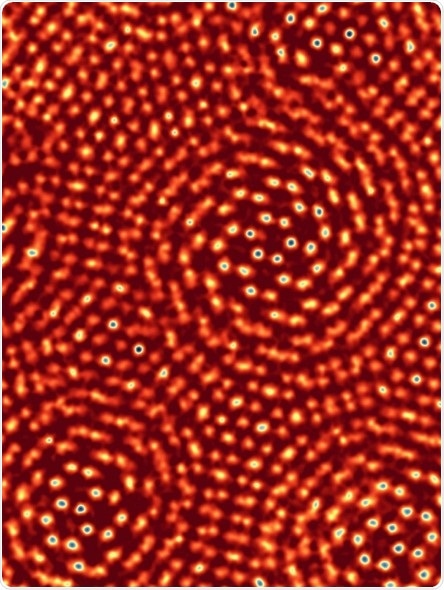
A ptychographic image of two sheets of molybdenum disulfide, with one rotated by 6.8 degrees with respect to the other. The distances between individual atoms range from a full atomic bond length down to complete overlap. Image courtesy of Prof. David Muller.
Ptychography is a computational imaging technique that utilizes sophisticated algorithms to reconstruct an image from overlapping diffraction patterns, created when an object is illuminated. In their paper, Muller’s group use a powerful new electron camera, which they have developed, to record the diffraction patterns with exceptional sensitivity, speed and dynamic range. The world-record resolution was made possible by the combination of detector and ptychography, heralding a new era in electron microscopy.
Phasefocus™ has commercialized ptychography as its proprietary platform technology, creating a portfolio of products to accommodate a wide range of imaging applications, encompassing life science, healthcare, engineering, metrology and more.
The Phasefocus πbox reconstruction engine, suitable for electron microscopy, is a back-end processing platform that can be connected to a microscope, network or cloud based, to deliver reconstructed ptychographic images to the user’s own software.
Compatible with electrons and any electromagnetic wave, πbox deciphers raw diffraction patterns to streamline image generation, making it applicable across a wide range of imaging modalities including X-ray, Electron microscopy and optical microscopy.
 Livecyte® Kinetic Cytometer system by Phasefocus wins 2017 Microscopy Today Innovation Award