Coronavirus disease 2019 (COVID-19) first arose in China, before spreading to nearly every country in the world. It has led to multiple health and economic crises, and the overall death toll is above six million.
 Study: Genetically engineered MRI-trackable extracellular vesicles as SARS-CoV-2 mimetics for mapping ACE2 binding in vivo. Image Credit: Volodymyr Dvornyk/Shutterstock
As would be expected, COVID-19 has been the focus of much research, both in search of treatments and vaccines and to better understand severe acute respiratory syndrome coronavirus 2 (SARS-CoV-2), the causative organism.
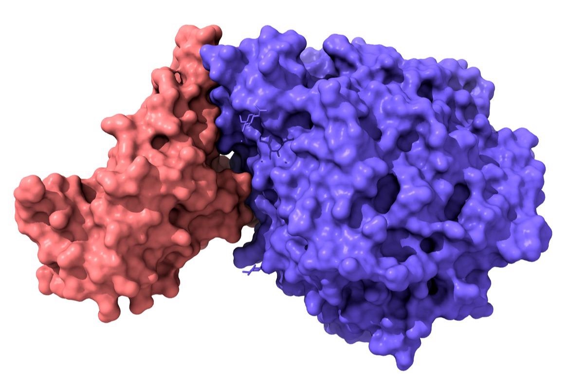
Now, researchers from the Weizmann Institute of Science have developed a method for tracking the binding of the receptor-binding domain (RBD) of SARS-CoV-2 in vivo using genetically engineered extracellular vesicles that can migrate to angiotensin-converting enzyme 2 (ACE2) expressing cells.
The study
A new pAGDisplay was created through three-component assembly of fragments from pDisplay, pLVX-Puro and pET26b plasmids in order to better engineer the pDisplay vector for efficient expression of viral peptides on the surface of extracellular vesicles (EVs) – specifically to create SARS-CoV-2 mimetics. The created plasmid used the puromycin resistance marker for increased ability to select, an IRES sequence downstream of the transmembrane domain to allow co-expression of the antibiotic resistance and transmembrane domain under a single promoter and the fluorescent protein eUnaG2 fused with the C-terminus of PuroR – allowing transfected cells to be easily detected.
As well as this, a RFnano protein was introduced to allow a more efficient selection of RBD-expressing cells using fluorescent activated cell sorting (FACS). The RBD of the original Wuhan strain was cloned at the N-terminus, and then specific tags were added to the constructs for further confirmation of expression.
Once the constructs were created, human embryonic kidney 293 (HEK293) cells were then transfected with pAGDisplay-RBD or pAGDisplay-noRBD, with FACS then used to select the cells with the highest expression levels of either eUNAG2 or RFnano.
Puromycin antibiotic selection was continued for three weeks to create stable cell lines expressing RBD. The stable cell lines were then characterized using immunostaining, immunoblotting, flow cytometry, and confocal microscopy.
The presence of RBD fused to the ALFA tag on the cell surface was confirmed, as were RBD-expressing cells incubated with the antibody – which showed bright signals on the cell surface corresponding only to the presence of RBD cells. It was also confirmed using flow cytometry that an anti-ALFA tag nanobody would only bind to the RBD cells. The recombinant extracellular portion of the ACE2 protein was then purified and conjugated to a fluorophore before incubation with noRBD or RBD cells. Fluorescence microscopy confirmed that the ACE2 protein was bound only to RBD cells.
After the validation of the expression of functional RBD on the surface of the cells secreted EVs were obtained using differential centrifugation. The EVs recovered were called EVrbd or EVnorbd respectively and were further examined using Western blot analysis which confirmed the expression of RBD through detection of the ALFA-tag in only EVrbds. To examine the binding capabilities of the EVs to ACE2 expressing HEK cells as opposed to control HEK cells, both types were then incubated with the labeled EVs for three hours before examination with fluorescence microscopy and flow cytometry. This revealed clear red fluorescence in HEK-ACE2 cells, but not in control HEK cells.
Following these successes, the researchers moved to in vivo experiments, examining the targetability of the EVrbds towards ACE2 receptors in immunodeficient mice implanted with HEK293 or ACE2-HEK293 cells. Two weeks after this implantation, the mice were intravenously injected with fluorescently labeled EVs. Six hours after this procedure, the implanted cells were excised and the fluorescent signal was analyzed, revealing a significantly higher accumulation of the EVrbds in the ACE2-HEK293 cells.
Discussion and Conclusion
The authors highlight that they have successfully created a construct that allows HEK293 cells to produce extracellular vesicles containing the RBD protein which can be harvested and bound to fluorescent proteins. These EVs can then be used to track which cells are expressing ACE2 and have been shown to successfully migrate to ACE2 expressing cells within a mouse model.
In essence, these EVs can act as SARS-CoV-2 mimetics, allowing researchers to study virus receptor interactions within organisms. This could help inform the researchers investigating SARS-CoV-2 and might potentially help treatments to be developed.
As well as this, the same approach could be used to investigate the role of other viruses in vivo.