New features enable scalability for all researchers with manual or automated workflows
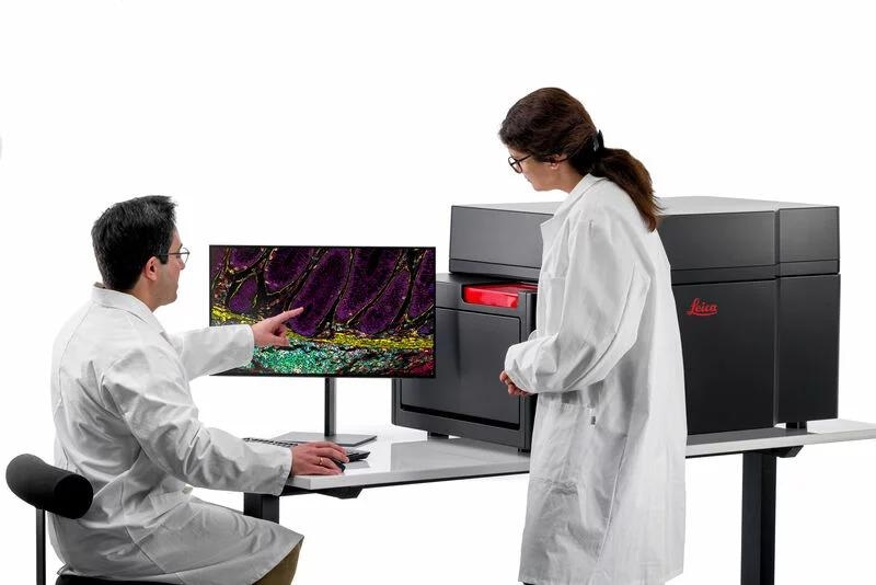
Image credit: Leica Microsystems GmbH
7 June 2022, Wetzlar, Germany – Leica Microsystems, a leading provider of microscopy and scientific instruments, has introduced a new generation of its multiplexed imaging solution, Cell DIVE, including software and hardware improvements. The more scalable and efficient multiplexing platform addresses spatial cell biology and function within the tissue microenvironment, offering researchers the freedom to select from over 350 rigorously validated antibodies to design their studies in their own way and a proven workflow to image 60+ biomarkers in a single tissue section.
The new Cell DIVE ClickWell coverslip-free slide holder enables users to automate their workflows based on their unique needs. The improved Cell DIVE workflow reduces the risk of unnecessary tissue damage and improves sample processing time while still offering the researcher the freedom to choose the staining method that is best for their research.
“Cell DIVE enables scientists, particularly those working in cancer research, to map normal and diseased tissue by cell type, biomarker profile, and specific features better than ever before,” says James O’Brien, Vice President of Life Sciences at Leica Microsystems. “Cell DIVE with ClickWell powers open multiplexing by making the manual workflow more efficient. It also gives researchers the power to choose their automation strategy to scale their research as it suits them.”
Cell DIVE sample staining occurs off the imager, allowing the user to process many slides through staining and dye inactivation steps in batches, thereby enabling scalability even without automation. ClickWell provides flexibility for automating sample staining, enabling the use of automated pipetting systems and autostainers. Researchers can also add a robotic arm to support unattended imaging of many samples.
The Cell DIVE platform was developed for scientists by scientists, enabling life science researchers to build accurate maps of the cellular architecture of healthy and diseased tissues, providing a deeper understanding of the human immune system, using credible data. The technology has been used in over 40 peer-reviewed publications, helping researchers deepen their understanding of the tissue microenvironment by offering outstanding spatial mapping of single cells within context.
About Leica Microsystems
Leica Microsystems develops and manufactures microscopes and scientific instruments for the analysis of microstructures and nanostructures. Ever since the company started as a family business in the nineteenth century, its instruments have been widely recognized for their optical precision and innovative technology. It is one of the market leaders in compound and stereo microscopy, digital microscopy, confocal laser scanning microscopy with related imaging systems, electron microscopy sample preparation, and surgical microscopes.
Leica Microsystems has six major plants and product development sites around the world. The company is represented in over 100 countries, has sales and service organizations in 20 countries, and an international network of distribution partners. Its headquarters are located in Wetzlar, Germany.
 Exploring electron microscopy for plant and microbial imaging at the John Innes Centre