From gait speed and DNA methylation to AI scans and clinical text, a major review maps how scientists are measuring aging, and why the field still needs stronger evidence before these tools guide patient care.
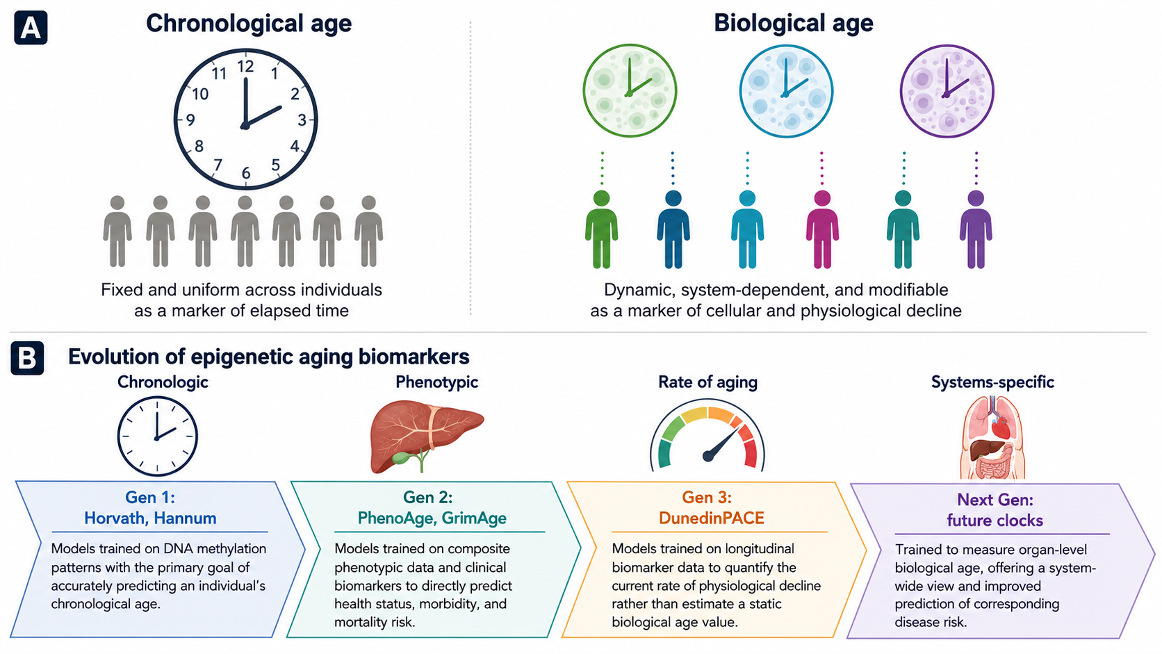
The distinction between CA and BA and the progression of epigenetic clocks through multiple generations. (A) Chronological age reflects a uniform measure of time, whereas biological age reflects cellular and physiological decline. (B) First-generation clocks (e.g., Horvath, Hannum) were trained to predict chronological age. Second-generation clocks (e.g., PhenoAge, GrimAge) focus on phenotypic health and mortality risk. Third-generation clocks (e.g., DunedinPACE) measure the current rate of biological aging. The next generation of clocks aims to measure organ-specific biological age. Image adapted from Figure 1 in Cheema, B. S. et al. Functional, molecular, and digital measurements of biological age. The Journal of Clinical Investigation. 2026;136(12):e205777. doi:10.1172/JCI205777. Licensed under CC BY 4.0.
In a recent review published in The Journal of Clinical Investigation, researchers discuss advances in biological age (BA) estimation over the years and ongoing limitations that need to be addressed for future clinical translation.
Pending prospective validation, clinicians may eventually use these approaches to identify high-risk individuals and support more personalized prevention or treatment strategies, but their role in routine care remains unresolved.
People of the same sex, born in the same year, with similar clinical profiles, such as adiposity, can differ in their physical abilities, immune resilience, likelihood of developing disease, and overall survival. This reflects cumulative effects of aging shaped by environmental exposures, health behaviors, genetic predisposition, molecular changes, and chance events. Biological age (BA) can capture such individual-level variabilities.
Scientists are developing new strategies to determine the biological age of different cells, tissues, and organs. Such efforts could support precision medicine approaches and ultimately improve the standard of care.
In this review, researchers provide an overview of BA estimation approaches and the ongoing challenges and limitations.
Conventional and modern methods for biological age estimation
Early-age estimation models primarily used predefined variables, risk factors, and functional markers (such as gait speed and grip strength), and interpreted the results as delayed or accelerated aging. They also developed composite scores for physiological aging. Although these methods help estimate disease risk, they are usually unable to explain cellular-level differences that may influence mobility, strength, and balance, which can affect the ability to perform daily tasks.
Researchers are now integrating molecular profiling with artificial intelligence (AI) algorithms into unified frameworks to improve BA estimation. First-generation clocks, such as Horvath and Hannum, used supervised machine learning models based on selected CpG methylation sites to estimate chronological age, whereas later clocks were designed to better capture phenotypic aging, morbidity, and mortality risk.
Digital AI models can analyze imaging, electrophysiology, wearable signals, and clinical text, while large language model (LLM)-based approaches have shown promise for estimating phenotypic aging from routinely collected health examination reports and electronic health records.
Decreases in gait speed and grip strength are associated with poorer physical function and higher mortality risks. New models are using video-derived and computer-based analyses to make these assessments more scalable.
As people grow older, the inner lining of the arteries becomes less efficient. Flow-mediated dilation provides an ultrasound-based window into endothelial function, while pulse-wave velocity and pulse pressure capture related aspects of arterial stiffness and vascular aging.
Wearables and digital monitors may contribute continuous data on heart rate, activity, sleep, and related physiological patterns, while ECG-derived age, inflammatory biomarkers, and measures such as maximal oxygen uptake provide complementary windows into cardiovascular and whole-body physiological reserve.
Elevated levels of natriuretic peptides such as NT-proBNP can help estimate long-term cardiovascular disease risk at levels far below standard measures. Likewise, cystatin C, a renal marker, has outperformed traditional creatinine-based indices in predicting mortality risks.
Deep learning models can estimate BA from brain magnetic resonance imaging (MRI) scans and high-resolution retinal blood vessel images. Advanced ML models can also predict survival outcomes in cancer patients using facial imaging, while molecular profiling can capture age-related biological patterns that may improve risk stratification. For instance, higher PAI-1 levels in midlife women have been associated with adverse metabolic and cardiovascular profiles.
BA estimation using composite approaches such as the Klemera–Doubal method and PhenoAge, as well as DNA methylation clocks such as GrimAge and DunedinPACE, has improved mortality risk determination by combining clinical biomarkers, biological processes, and methylation-based aging signals.
New clocks, such as Systems Age, OMICmAge, GlycanAge, and MileAge, can quantify changes across multiple organ systems, immune cell populations, and metabolic pathways. Proteomic aging clocks (PAC) can now analyze thousands of proteins using regression models to help estimate BA, health span, disease susceptibility, and mortality risk.
Challenges and Future Directions
The scientific community has made significant strides in age estimation. Nevertheless, a few challenges remain. There is no standardized, unified framework for determining an adequate BA measure. Moreover, most BA models rely on cross-sectional analyses and prognostic information.
Head-to-head comparisons of methodological reliability, response to treatments, feasibility, and long-term stability are required to understand whether changes in BA measurements reflect real improvements or declines in health.
Scientists must also improve AI models to address information transparency and delineate age-related changes from disease pathophysiology for biomedical applications. Because predictive association does not prove causal modifiability, researchers must also determine whether lowering a BA estimate corresponds to meaningful improvements in health span, function, or survival. Coordinated interdisciplinary efforts and increased investment in healthcare services to link BA measures with clinical outcomes will help close these gaps.
Prospective studies including human participants and unified testing measures are required to accelerate the clinical translation of BA measures.
Conclusions
Based on the findings, advanced biological age estimation methods generally show strong predictive promise compared with traditional approaches. New frameworks use AI and machine learning to analyze multi-modal data, including medical records, lab test values, clinical findings, imaging results, and genetic information.
These integrated frameworks may capture complementary dimensions of aging beyond traditional single-modality approaches, but direct comparative and longitudinal evidence remains limited. However, further research is needed to link BA estimates with clinical findings for application in real-world settings.
Download your PDF copy by clicking here.
Journal reference:
- Cheema, B. et al. (2026). Functional, molecular, and digital measurements of biological age, The Journal of Clinical Investigation, 136(12):e205777. DOI: 10.1172/JCI205777, https://www.jci.org/articles/view/205777
 Single-dose gene therapy extends healthy lifespan in older mice