What happens when the gut loses its natural hormonal defenses against immune attack, and could existing medicines offer a new approach to protecting the intestinal lining?
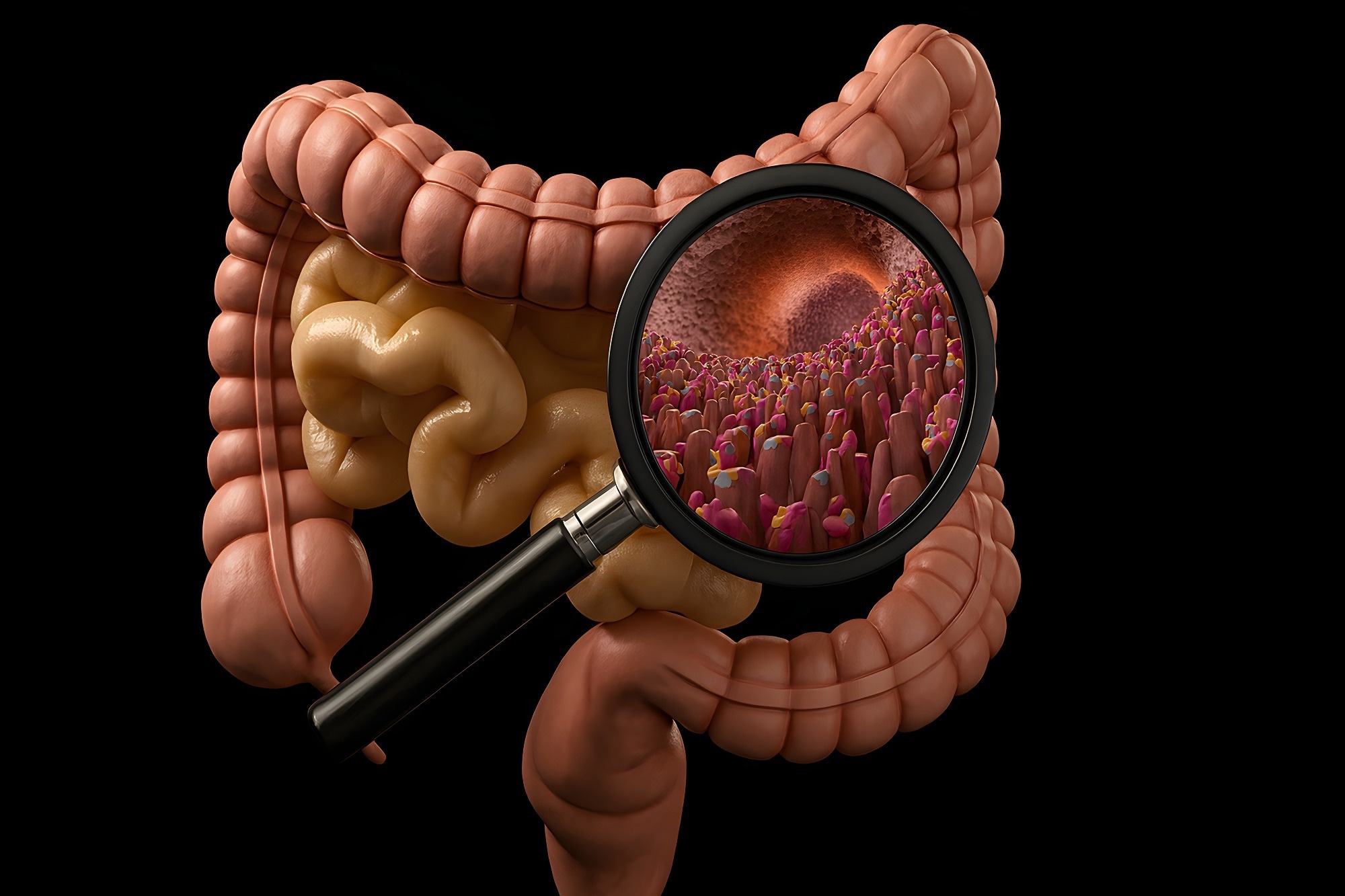
Study: Gut-derived glucagon-like peptides attenuate T cell-induced intestinal injury. Image Credit: DRMEK / Shutterstock
A recent study published in the journal Cell Reports Medicine investigated how gut-derived glucagon-like peptides affect intestinal damage induced by activated T cells. In mice, signaling through the glucagon-like peptide-1 and peptide-2 receptors (GLP-1R and GLP-2R) helped limit this damage. Mice lacking proglucagon expression in the intestine were more susceptible to injury. Semaglutide and teduglutide, which activate GLP-1R and GLP-2R, respectively, reduced intestinal damage caused by CD3ε-targeting antibodies. These findings suggest that gut-derived GLP hormones may help protect the intestine during acute inflammation.
Specialized L cells in the gut release GLP hormones, which serve different functions. While GLP-1 helps regulate blood glucose and appetite, GLP-2 supports nutrient absorption, intestinal lining growth, barrier integrity, and blood flow. Animal studies have also found that treatment with agonists of these receptors can reduce gut damage, prompting further investigation of their potential protective effects.
About the study
In the present study, the researchers examined whether activating GLP hormone receptors could help protect the gut from T cell-induced damage in mice.
To examine the role of gut-derived glucagon-like peptides, the researchers used mice lacking GLP-1R or GLP-2R, alongside animals with proglucagon expression selectively removed from the intestine (GcgGut−/−). The experiments used male mice aged 12 to 16 weeks. They also tested semaglutide at 2.5 μg/kg and teduglutide at 40 μg/kg to determine whether activating these receptors could limit intestinal injury. Separate immune-cell experiments used a higher semaglutide dose of 10 μg/kg. 16S ribosomal RNA sequencing characterized changes in the fecal microbiome, with alpha diversity assessed using the Shannon and Simpson indices and microbial richness estimated using Chao1.
After anti-CD3 treatment, immune cells from the mesenteric lymph nodes (MLNs), small intestinal lamina propria (LP), and spleen were examined by flow cytometry. The team also measured mRNA levels of the intestinal stem cell markers olfactomedin 4 (Olfm4) and leucine-rich repeat-containing G protein-coupled receptor 5 (Lgr5). Other measurements included lipocalin 2 (Ngal), the Paneth cell markers lysozyme 1 (Lyz1) and defensin alpha 5 (Defa5), and inflammatory cytokines. The researchers also examined crypt density and Paneth cell loss to assess intestinal damage.
Results and discussion
T cell-induced intestinal damage was increased in Glp1r−/−, Glp2r−/−, and GcgGut−/− animals, but the abnormalities differed across models. GLP-1R deficiency increased inflammation and histological damage without increasing crypt loss compared with controls. GLP-2R deficiency reduced villus height and crypt cell proliferation and increased Paneth cell loss without reducing small-bowel weight or length. Mice lacking intestinal proglucagon developed greater intestinal permeability, more extensive crypt loss, and reduced small-bowel weight. In mice lacking GLP-1 receptors (Glp1r−/−), the researchers found higher ileal lipocalin 2 (Ngal) gene expression and concentrations of C-X-C motif chemokine ligand 1 (CXCL1), IL-1β, and IL-6. The number of neutrophil elastase (NE)-positive cells was also elevated. By comparison, the expression of Niemann-Pick C1-like 1 (Npc1l1), intestinal fatty acid-binding protein 2 (Fabp2), and regenerating islet-derived protein 3 gamma (Reg3g) was reduced in Glp1r−/− animals. Together, these findings suggest a heightened inflammatory response in these animals.
GLP-1 levels were considerably lower in proglucagon-deficient mice. In these animals, CXCL2 expression increased, while the intestinal villi were shorter. The researchers also detected lower ileal mRNA levels of Slc10a2, Olfm4, and Lgr5. The findings were accompanied by delayed body weight (BW) recovery following anti-CD3 challenge. Anti-CD3 treatment reduced microbial richness in both genotypes, while Shannon and Simpson indices indicated lower diversity in intestinal proglucagon-deficient mice than in controls. Following T cell activation, Verrucomicrobiota and Proteobacteria made up a larger share of the gut microbiota, whereas Firmicutes became less abundant in both genotypes. T cell activation, rather than intestinal proglucagon deficiency, accounted for most changes in microbial composition.
Systemic T cell activation induced intestinal GLP-1 and GLP-2 secretion, and endogenous GLP-1R/GLP-2R signaling protected against T cell-induced intestinal injury. Treatment with semaglutide or teduglutide reduced anti-CD3-induced intestinal injury, with combination treatment also showing protective effects.
Teduglutide partly restored the small-bowel weight-to-length ratio, reduced intestinal cytokine levels and neutrophil infiltration, and eased tissue damage, but did not improve measured indicators of intestinal permeability. Semaglutide lowered cytokine concentrations in the blood and increased the expression of Paneth cell-related genes in mice, with selective removal of intestinal proglucagon expression following anti-CD3ε antibody treatment. Combination therapy lowered systemic inflammatory markers, restored ileal crypt density, and reduced histological damage. Although both drugs showed protective effects, the findings did not establish whether combining them offered additional benefits over either treatment alone.
Semaglutide also reduced blood glucose excursions during an oral glucose tolerance test (OGTT) and increased the expression of genes associated with Paneth cells and intestinal epithelial activity. Paneth cell-related gene expression rose in proglucagon-deficient mice, while Fgfbp1 increased more prominently in controls and Fabp2 increased only in controls. Combination therapy increased Ereg and Egr1 gene expression.
Flow cytometry found largely unchanged immune cell proportions in the intestinal LP and MLNs after anti-CD3 treatment, while splenic T cell proportions were lower in mice lacking intestinal proglucagon. Semaglutide and teduglutide caused few changes in immune cell subset proportions.
Earlier studies using models of colitis and mucositis have reported that GLP-1R agonists may help ease gut inflammation and stimulate mucus production by Brunner’s glands. GLP-2, meanwhile, supports the intestinal lining by promoting cell growth and limiting cell death. Animal studies also suggest that mice lacking GLP-2R are more prone to Paneth cell dysfunction and enteritis. Beyond these effects, GLP-2 may influence group 2 innate lymphoid cells, while GLP-1 can encourage intestinal growth through crypt fission. These actions may vary with the type of intestinal injury.
Conclusions
The findings suggest that endogenous GLP-1R and GLP-2R signaling protects against T cell-induced mucosal damage, while pharmacological activation of these receptors can reduce intestinal injury. Loss of intestinal proglucagon expression increased susceptibility to T cell enteropathy in mice.
The experiments were short and involved young male mice with acute intestinal injury, so the benefits cannot yet be assumed to apply to human disease. Intestinal proglucagon deletion also reduces other related peptides, which may have contributed to the findings.
In future studies, researchers could investigate whether lower doses of GLP-1R agonists or combined GLP-1R and GLP-2R agonism could reduce immune-mediated gut injury without causing considerable weight loss. Researchers could also examine the roles of other glucagon-derived peptides, including glicentin and oxyntomodulin, and assess longer treatment regimens and different agonist doses in mice with diabetes or obesity.
Source:
Journal reference:
 GLP-1 treatment linked to 40% fewer asthma attacks