Two people with equally sharp memories may face very different cognitive futures. Scientists have uncovered a brain connectivity signature that could reveal who is most vulnerable to Alzheimer’s disease and who is more likely to maintain their cognitive abilities as they age.
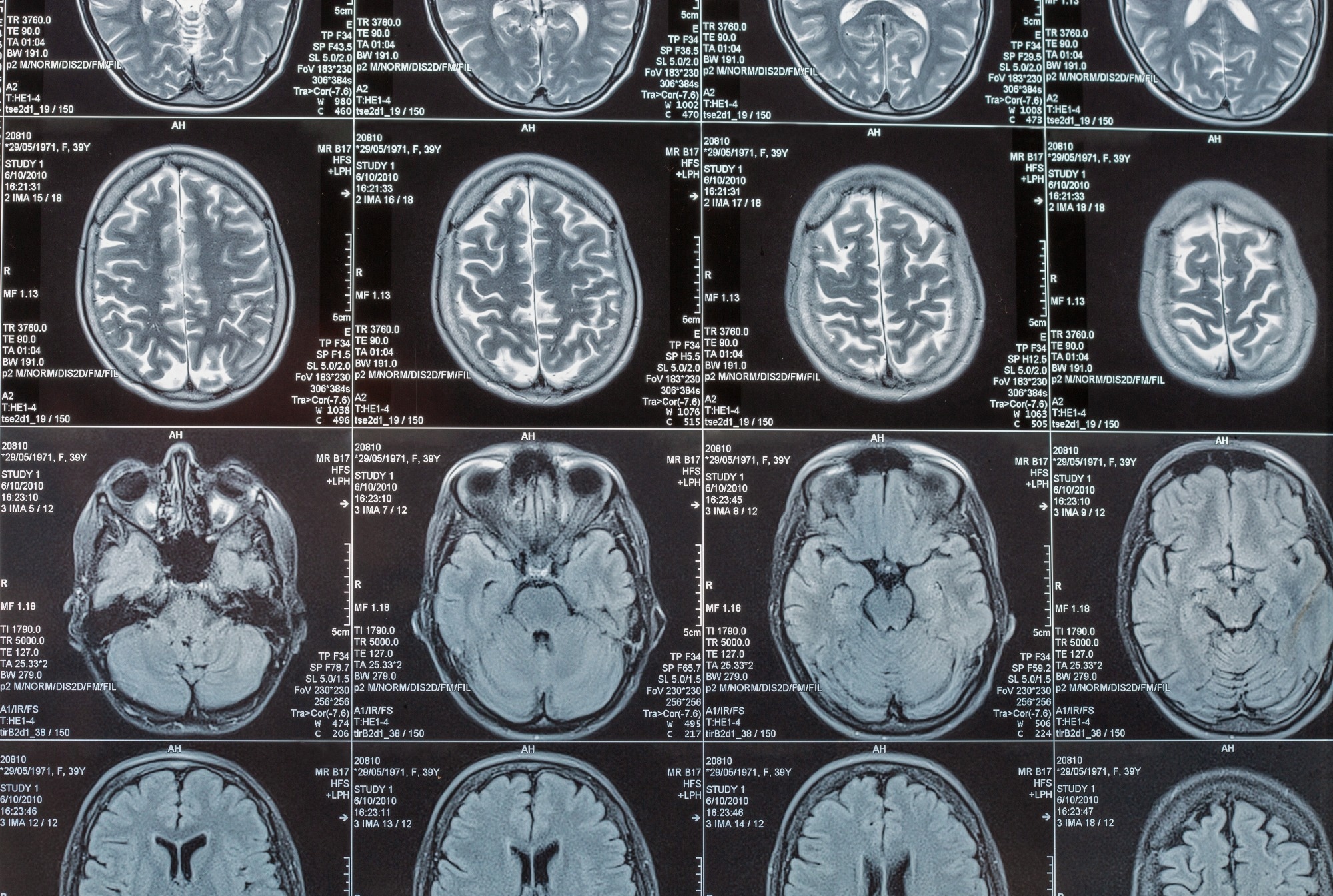 Study: A functional connectome identifies vulnerability to cognitive decline and Alzheimer’s disease progression. Image credit: Sergiy Palamarchuk/Shutterstock.com
A new study published in Science Advances identifies distinct brain connectivity patterns that may help predict the risk of cognitive decline and progression to Alzheimer’s disease before clinical symptoms appear.
How brain connectivity distinguishes superagers from Alzheimer’s patients
Brain aging is a natural consequence of the physiological aging process, characterized by gradual structural, cognitive, neuronal, and chemical changes in the brain over time. Not everyone experiences these changes at the same rate. Some people experience more rapid brain aging, especially those with a disease like Alzheimer’s disease-related dementia. In contrast, some people, termed “Superagers”, maintain youthful cognitive performance in their old age.
Characterization of the neurobiological basis of these divergent brain aging dynamics and identification of the specific brain connectivity patterns that can resolve this heterogeneity may be crucial for predicting future risk of cognitive decline and Alzheimer’s disease and related dementias.
Focusing on this important research area, a research team led by Stanford Medicine analyzed resting-state functional magnetic resonance imaging (fMRI) scans obtained from 180 superagers and 110 age-matched adults diagnosed with Alzheimer’s disease. Superagers were aged 75 years or older, with episodic memory performance exceeding that of middle-aged adults and executive function comparable to that of their age-matched peers.
They derived the Alzheimer’s Resilience-Vulnerability Connectome (ARC) by comparing fMRI scans of superagers and those with Alzheimer’s disease. The identified brain connectivity signature involved prefrontal, temporal, insular, and parietal regions associated with memory, executive function, and emotional regulation. The score effectively distinguished these two subsets of individuals with approximately 80% accuracy in the discovery cohort and 71% accuracy in an independent replication cohort of 143 individuals.
Brain connectivity predicts cognitive decline before symptoms appear
The researchers subsequently evaluated the ARC score in 1,315 cognitively unimpaired adults across a discovery cohort of 818 individuals and an independent replication cohort of 497 individuals. ARC-based subtyping in this population led to the identification of superager-like and Alzheimer’s disease-like subgroups.
Both subgroups exhibited similar cognitive performance and comparable amyloid-beta and tau burdens at baseline but divergent clinical outcomes over the longitudinal follow-up period. People with Alzheimer’s disease-like brain connectivity patterns exhibited faster cognitive decline, more rapid accumulation of amyloid-beta plaques (primary biomarkers of Alzheimer’s disease), and higher risk of progression to mild cognitive impairment or Alzheimer’s disease during follow-up. In contrast, people with superager-like brain connectivity patterns generally maintained stronger cognitive abilities.
The Alzheimer’s disease-like subgroup also showed signs of accelerated functional brain aging. ARC scores were more closely associated with the estimated biological age of the brain than with an individual’s chronological age.
Although individuals in the Alzheimer’s disease-like subgroup were older at baseline, differences in subsequent cognitive decline persisted after accounting for baseline age differences.
ARC reveals cognitive vulnerability beyond amyloid and tau
Amyloid-beta and tau proteins are major biomarkers associated with the development and progression of Alzheimer’s disease. The disease is characterized by amyloid plaques outside nerve cells and tau tangles within them. However, although these biomarkers can be detected before clinical symptoms appear, they do not fully explain why some individuals experience more rapid cognitive decline than others.
The researchers explored the usefulness of the ARC score as an early marker for detecting signs of cognitive decline. Such a marker can potentially help identify at-risk people and inform future preventive strategies.
The analysis showed significant associations between ARC scores and amyloid-beta burden across the clinical spectrum. However, associations with tau pathology were less consistent, and the two subgroups did not differ significantly in their rates of tau accumulation. Overall, the ARC score provided additional predictive information beyond conventional biomarkers in assessing the risk of progression to mild cognitive impairment or Alzheimer’s disease, while offering information at an early stage. The integration of ARC scores with conventional biomarkers significantly improved the prediction.
Notably, the ARC score helped distinguish individuals with different rates of cognitive decline even among those who already had amyloid pathology, independently of their baseline clinical function and amyloid-beta and tau burdens.
The ARC scores also exhibited significant associations with daily functional impairment and memory decline in cognitively unimpaired adults. These findings highlight the relevance of ARC beyond the two original groups of superagers and individuals with Alzheimer’s disease and within cognitively unimpaired adults.
Brain connectivity offers new opportunities for early risk assessment
The researchers established a reproducible brain connectivity signature (ARC) that can distinguish between cognitively unimpaired older adults who are more likely to experience cognitive decline and progress into Alzheimer’s disease and those who are more likely to maintain their cognitive abilities as they age.
Rather than simply separating individuals into two groups, ARC captures a continuous spectrum of cognitive aging, with cognitively average older adults occupying an intermediate position between superagers and individuals with Alzheimer’s disease.
As observed in the study, these signatures provide additional predictive information beyond conventional Alzheimer’s disease biomarkers and genetic risk factors in predicting future cognitive decline. Taken together, these findings support the potential utility of ARC for early risk stratification among cognitively unimpaired older adults. However, further prospective validation of these findings against established and emerging Alzheimer’s disease biomarkers is required to establish its clinical utility.
The exploratory genome-wide association study conducted by the researchers identified three suggestive genetic variants near CLYBL and FRMD6 associated with differences between ARC-defined Alzheimer’s-like and superager-like subtypes.
Existing evidence links CLYBL to citrate metabolism and inflammatory pathways and FRMD6 to neuronal morphogenesis and synaptic architecture. These observations suggest a potential link between molecular variation and system-level brain organization. However, the current study findings did not reach conventional genome-wide significance and were not independently replicated. Future larger-scale genetic studies are therefore required to determine whether these loci reproducibly contribute to individual differences in Alzheimer’s disease-related brain aging trajectories.
The researchers relied exclusively on resting-state fMRI to characterize brain connectivity, raising the possibility that they missed other critical neurobiological features. Integrating multimodal neuroimaging measures, such as structural connectivity and structure-function coupling, may offer additional understanding of aging-related brain changes.
Given the observational nature of the study, the researchers could not determine whether ARC specifically reflects the brain’s ability to maintain cognitive function despite existing Alzheimer’s disease pathology or its ability to resist the accumulation of such pathology. Further studies involving more ethnically diverse populations and using established definitions of superagers are also needed to confirm the generalizability of the findings.
The researchers did not model other common aging pathologies, such as vascular contributions to cognitive decline. The ARC score should therefore be interpreted within an Alzheimer’s disease-related framework.
 Could eating more mushrooms help protect your brain as you age?