Over the last 15 years, functional magnetic resonance imaging (fMRI) of the brain's "resting state" (rsfMRI) and its measures of functional connectivity have been major topics of interest.
It is assumed that low-frequency fluctuations seen in the BOLD signal reflect sudden neural activity and that synchronized fluctuations in separate brain regions thus point to a functional link between them, as illustrated in figure 1.
Figure 1. Principle of Seed based Functional Connectivity Analysis. Image Credit: Bruker BioSpin Group
Initially demonstrated for the human motorcortex (Biswal et al., 1995), a number of connectivity networks have been discovered in the human brain and have been shown to vary in different psychiatric and neurological diseases. This makes functional connectivity MRI (fcMRI) a highly interesting method to improve our understanding of brain function with regards to health and disease.
fcMRI Technique
The establishment of fcMRI in animal models was started only a few years ago. The technique is very attractive, especially in rodents, as it is anticipated to offer a highly interesting functional readout for disease progression, treatment and repair in a wide range of existing animal models.
When compared to fMRI, fcMRI does not depend on stimulation and can explore brain networks that cannot be accessed via external stimuli. It also leverages the major benefits of MRI with regard to non-invasiveness and suitability for longitudinal studies.
This technical brief assesses the feasibility and potential of fcMRI applied to imaging of rat brain through a high field MRI system.
Experimental Framework
MRI System
117/16 Bruker BioSpec ('H @ = 500 MHz); BGA9s gradient system (Gmax=750 mT/m; min. ramp time of 125 µs); quadrature volume resonator (inner diameter 72 mm) for transmission; Avance II electronics; ParaVision; rat brain quadrature surface coil (-30x30 mm2) for reception.
Physiological Monitoring
Monitoring system (SA Instruments): fiber optic temperature probe, pulse oxymeter, display and recording (synced to MRI acquisition) of respiration, respiratory pad, pulse and temperature utilizing a custom-made data acquisition software (DasyLab).
Imaging Protocol
FieldMap acquisition and local MapShim; TurboRARE for T-weighted anatomical reference (RARE-factor 8, 28 slices a 0.5 mm, 2562 matrix, 125 x 125 µm2, TR 4.0 s, TEoff 32.5 ms, 2 averages, acq. time 4m16s); single-shot gradient echo EPI for functional image acquisition.
Source: Bruker BioSpin Group
Anaesthesia Protocol
Initial anaesthesia utilizing 1.5 % Iso-flurane; conversation to Medetomidine sedation: subcutaneous 0.5 ml bolus and ensuing infusion of 1 ml/h of Domitor solution (Pfizer; 0.1 ml/kg ad 10 ml saline solution).
Processing and Analysis
Motion correction and coregistration to rodent brain template utilizing FSL tools; connectivity analysis with custom-made ImageJ Software and FSL tools; removal of physiological noise utilizing regression of motion and physiological parameters (Kalthoff et al., 2011).
Results and Discussion
High field BOLD functional MRI information of the resting rat brain consistently attains a quality expedient for functional connectivity analysis.
Using the given EPI imaging protocol, voxel time courses generally have signal variations on the order of 1.5% that occur from a number of sources, as shown in figure 2.
Figure 2. Contributions to Resting State BOLD Signal Fluctuations. Image Credit: Bruker BioSpin Group
One third of the fluctuations are attributed to the system’s intrinsic raw noise, mainly thermal noise from coil, sample and electronics. Most (~40%) of the fluctuations occur as a result of physiological noise that begin from the cardio-respiratory cycle and related pseudo-motion.
To reduce false-positive correlations in ensuing analyses, physiological noise should be rectified by motion correction and regression, which can enhance tSNR by ~30%. The remaining ~25% of fluctuations may be due to actual neurovascular fluctuations at which the succeeding functional connectivity analysis is aimed.
Seed-based connectivity analysis (SCA) can produce connectivity maps that are able to visualize the temporal correlation of voxels with seed regions defined by priori. Rodent brain functional connectivity maps from seed regions in striatum and cortex reveal clear bilateral regions, suggesting powerful interhemispheric connectivity between homologous regions, as illustrated in figure 3.
It should be noted that observation of reliable, robust and particular connectivity networks may not be possible through traditional Isoflurane anaesthesia, and needs highly sensitive experimental protocols like Medetomidine sedation (Weber 2006, Pawela 2008, Williams 2010).
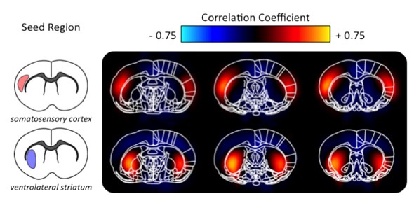
Figure 3. Seed based Functional Connectivity Maps (group average). Image Credit: Bruker BioSpin Group
In addition, the topology of functional connectivity networks can be studied utilizing data-driven methods such as independent component analysis (ICA). This analysis consistently detects separated brain functional networks in both cortical and subcortical structures on a per-subject basis, as indicated in figure 4. Such methods are especially interesting to compare network topologies without a priori hypotheses between species or in pathologies.
Figure 4. Topography of Functional Connectivity Networks identified via ICA. Image Credit: Bruker BioSpin Group
It has been identified from human studies that functional connectivity is susceptible to various neurological disorders. Initial studies demonstrate that this holds true for a number of animal models, for instance in the longitudinal evaluation of network remodelling after stroke, as demonstrated in figure 5.
Figure 5. Loss of Functional Connectivity after Stroke.Image Credit: Bruker BioSpin Group
Conclusion
Functional connectivity magnetic resonance imaging of the rat brain can deliver a functional readout for disease progression, treatment and repair in a number of existing animal models.
Through technical progression and extensive availability of high field systems, the challenge of fcMRI has moved towards novel data analysis strategies and maintenance of a suitable physiological state.
Functional connectivity, especially in tandem with other modalities, will be a key concept for neuroscientific research in the coming years. One of the crucial steps to manipulate this potential is to set up standard protocols for mouse fcMRI to access the wide range of transgenic animal models.
Acknowledgement
Produced from articles authored by Daniel Kalthoff and Mathias Hoehn.
Sources
- Biswal B, Yetkin FZ, Haughton VM, Hyde JS (1995) Functional connectivity in the motor cortex of resting human brain using echo-planar MRI. Magn Reson Med 34:537-41
- Weber R, Ramos-Cabrer P, Wiedermann D, van Camp N, Hoehn M (2006) A fully noninvasive and robust experimental protocol for longitudinal fMRI studies in the rat. Neuroimage 29:1303-10
- Kalthoff D, Seehafer JU, Po C, Wiedermann D, Hoehn M (2011) Functional connectivity in the rat at 11.7T: Impact of physiological noise in resting state fMRI. Neuroimage 54:2828-39
- Pawela CP, Biswal BB, Cho YR, Kao DS, Li R, Jones SR, Schulte ML, Matloub HS, Hudetz AG, Hyde JS (2008) Resting-state functional connectivity of the rat brain. Magnetic Resonance in Medicine 59:1021-9
- Williams KA, Magnuson M, Majeed W, LaConte SM, Peltier SJ, Hu X, Keilholz SD (2010) Comparison of alpha-chloralose, medetomidine and isoflurane anesthesia for functional connectivity mapping in the rat. Magn Reson Imaging 28:995-1003
- Beckmann CF, DeLuca M, Devlin JT, Smith SM (2005) Investigations into resting-state connectivity using independent component analysis. Philosophical Transactions of the Royal Society B-Biological Sciences 360:1001-13
- Jonckers E, Van Audekerke J, De Visscher G, Van der Linden A, Verhoye M (2011) Functional Connectivity fMRI of the Rodent Brain: Comparison of Functional Connectivity Networks in Rat and Mouse. PLoS One 6:e18876
About Bruker BioSpin Group
The Bruker BioSpin Group designs, manufactures, and distributes advanced scientific instruments based on magnetic resonance and preclinical imaging technologies. These include our industry-leading NMR and EPR spectrometers, as well as imaging systems utilizing MRI, PET, SPECT, CT, Optical and MPI modalities. The Group also offers integrated software solutions and automation tools to support digital transformation across research and quality control environments.
Bruker BioSpin’s customers in academic, government, industrial, and pharmaceutical sectors rely on these technologies to gain detailed insights into molecular structure, dynamics, and interactions. Our solutions play a key role in structural biology, drug discovery, disease research, metabolomics, and advanced materials analysis. Recent investments in lab automation, optical imaging, and contract research services further strengthen our ability to support evolving customer needs and enable scientific innovation.
Sponsored Content Policy: News-Medical.net publishes articles and related content that may be derived from sources where we have existing commercial relationships, provided such content adds value to the core editorial ethos of News-Medical.Net which is to educate and inform site visitors interested in medical research, science, medical devices and treatments.