The following piece is the final article in a three-part series on imaging large whole mount brain sections for neuroscience research. The first two articles covered sectioning and staining and scanning large brain tissue sections for neuroscience research. This article discusses data management in digital imaging of large tissue sections.
Introduction
Despite many advances in imaging technology and methodologies, neurohistology remains a key tool in neuroscience research. Recent developments in histotechnology capabilities mean it is now possible to image large tissue samples. This is of particular importance in the study of the brain since the connections between cells and different areas of the brain must be evaluated and related to sub-cellular structure.
Mapping the Brain Universe
Large tissue volumes must be visualized at high resolution to aid in the further understanding of the healthy brain and the changes that occur in neurological diseases. Although in vivo imaging can provide some valuable insights, it cannot supply the level of cellular detail obtained from histological imaging.
Sophisticated imaging hardware is now available that facilitates the imaging of large tissue samples. Automated imaging systems have significantly speeded up digital workflows, and the provision of digital slides has facilitated the sharing of histology research. However, systems for managing the data acquired are only just catching up.
With large tissue imaging producing a considerably greater volume of data than conventional slide imaging, the management of such quantities of data is challenging analysis software and data storage solutions. Having invested considerable time in preparing a large tissue section and acquiring high-quality, high-resolution images, it is necessary to ensure that adequate systems and workflows are in place to effectively manage the large volume of data generated.
This article highlights some of the data management challenges that arise from the imaging of large tissue samples.
Handling large amounts of imaging data
Digital images of large tissue samples have huge file sizes. Whereas images from the scanning of conventional slides will be measured in megabytes, scanning of large tissue sections necessitates vast computer memory and storage capacity.
For example, a 175 mm x 125 mm human brain section scanned at 40X resolution can result in an image file as large as one terabyte in size (for an uncompressed image). If multiple Z-stack planes of a single section are imaged, the digital image size of the single section can increase to the order of tens of terabytes.
Dr. Savvas Damaskinos, CTO, Huron Digital Pathology warns: "Large area scans require significant software resources in terms of computing power and memory allocation".
Working with such large files can also overwhelm software capabilities. As discussed in the previous article, specific workflows and sophisticated algorithms must be developed to set up a large area high resolution scan. In addition, specialized software for the graphical analysis of images from large tissue volumes is needed.
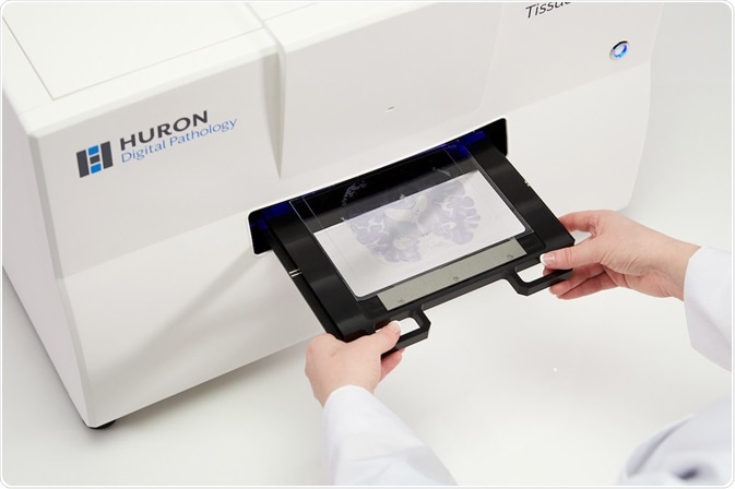
TissueScope LE Whole Slide Scanner. Image Credit: Huron Digital Pathology
Despite continual software improvement, the sizes of files are growing more quickly. Software issues aside, these analyses produce vast datasets that must be stored somewhere. Although there are companies who will produce and analyze digital images from large tissue slides, they do not offer a data storage facility.
Dr Robert Switzer, President and Chief Scientific Officer of NeuroScience Associates, explains: "The large files sizes are posing a challenge to many of the current software programs which have not grown to keep pace with the new capabilities of full-section digital capture. There are work-arounds, but the demand is there so it should improve in the near future—we hope!"
Storing large digital slides
The storage of data from large tissue imaging should be a key consideration in the development of any digital workflow. Forward thinking and systematic planning are fundamental to the success and cost-effectiveness of a digital workflow. The best storage solution can vary according to the precise nature of the workflow, so the whole process must be considered when choosing a storage solution.
Broadly speaking, there are two main options for storing digital slides: either on a dedicated server on the premises, or in cloud-based online storage. There are benefits of both and so, in order to choose the most appropriate option, it is important to determine how the digital images will reach the chosen storage facility and how they are likely to be used in the future.
Storing digital slides on the premises requires the purchase of network attached storage devices, which are essentially additional hard drives that can be used to store digital files. The initial investment will be offset by savings in the purchase of online storage space. However, you will also need to ensure suitable back-up to protect against damage or malfunction, and this may be riskier than using the online back-up services of a specialized company.
The additional time needed for creating such back-ups may well be offset by obviating the need to upload the files, which can be time consuming unless a good overall internet bandwidth is available. If remote access to the digital slide bank is likely to be needed, it is important to ensure that the available network supports this.
Cloud storage can be purchased from a range of suppliers, with the cost varying according to the amount of storage required. There may also be additional charges for moving items stored on the cloud or limits/charges imposed by the network for uploads. However, it is reassuring to know that everything uploaded to purchased online storage area will be automatically backed up by the provider so there is not the worry of losing the images obtained at great lengths.
As mentioned earlier, it is also worth bearing in mind that the upload process for large digital images may well be costly in terms of time as a result of bandwidth restrictions. This could produce a bottle neck in a digital workflow due to waiting times for images to be uploaded from the scanner. However, the main benefit is that access can be readily given to remote third parties.
Irrespective of the storage solution chosen, it is important that an effective catalogue system is designed and used so it is clear what is stored where and a particular image can be selected immediately at any point in the future. Effective storage systems should also ensure that a particular image is accessed from a single location and not saved to multiple local hard drives, whereby wasting valuable storage space as well as time.
W. Kemp Watson, CEO and Founder, Objective Pathology Services, advises: "the biggest challenge is figuring out the best way NOT to move them – one of the great features of digital slides is that they can be viewed right from where they were created".
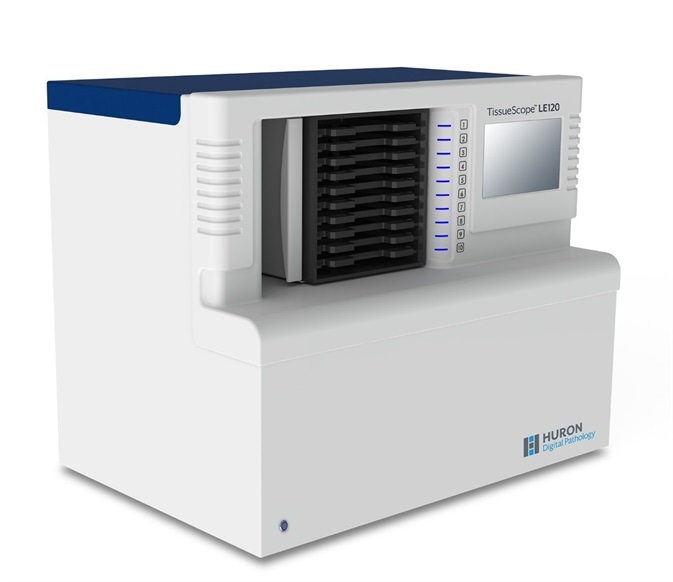
TissueScope LE120 Whole Slide Scanner. Image Credit: Huron Digital Pathology
Practical IT considerations
The practical implementation of digital workflows that include extremely large files is largely dependent on IT capabilities.
Digitized pathology images typically have very high resolution, which can make viewing the entire image on a computer screen difficult. Multiresolution “zooming” technology allows the entire original image to be viewed at lower resolution whilst allowing one-click access to full-resolution images of the areas of interest.
Increased capabilities of compression algorithms, such as JPEG 2000, have dramatically reduced the size of digital images, whereby significantly reducing storage costs.
However, despite this progress, there are still many areas in need of improvement. A key short-coming is the lack of standard file formats and application program interfaces. Consequently, software is not readily interchangeable. If a programmer has designed a software for imaging and analysis in accordance with the requirements of one operating system or other application, it may not be compatible with a different system.
Finally, although the importance of obtaining high-quality high-resolution digital images has been embraced, the importance of having a fast and reliable network is often understated. This can severely impact the efficiency with which these images can be managed. Furthermore, access to high-speed broadband internet is not routinely available to all institutions.
Summary
Having invested significant resources into acquiring high-quality high-resolution digital images from large tissue samples, it is important that they can be analyzed appropriately to ensure correct interpretation of experimental results.
In addition to the huge size of large-format digital images, analysis of these images creates a vast quantity of data. Both the images and the data must be stored, either on a local server or on the cloud. There are benefits and disadvantages of both options, which must be considered in relation to the precise nature of the digital workflow and the intended use of the image.
Although it is possible for digital images to be accessed directly from the point of their creation without moving them, the efficiency of doing so can be hampered by broadband capacity and network limitations.
The size of digital images has suddenly burgeoned and it is hoped that IT capabilities will soon catch up in order to support this valuable research tool.
References
About Huron Digital Pathology
 Based in Waterloo, Ontario, Canada, Huron Digital Pathology has a 20 year history designing sophisticated imaging instrumentation. Our end-to-end digital whole slide imaging solutions for digital pathology incorporate our award-winning TissueScope™ digital slide scanners; TissueView™ image viewing, sharing and management platform; and our workflow-enhancing accessories, which include our innovative TissueSnap™ preview scanning station.
Sponsored Content Policy: News-Medical.net publishes articles and related content that may be derived from sources where we have existing commercial relationships, provided such content adds value to the core editorial ethos of News-Medical.Net which is to educate and inform site visitors interested in medical research, science, medical devices and treatments.