Introduction
Definition and Key Features
Genetic and Molecular Basis
Clinical Presentation
Diagnosis
Prognosis and Outcomes
Management and Supportive Care
Research and Future Directions
Conclusions
Related video: What Is Bent Bone Dysplasia?
References
Further reading
Bent bone dysplasia (BBD) is a perinatal-lethal skeletal disorder caused by pathogenic FGFR2 mutations that disrupt bone formation, leading to bent long bones, osteopenia, and craniosynostosis.
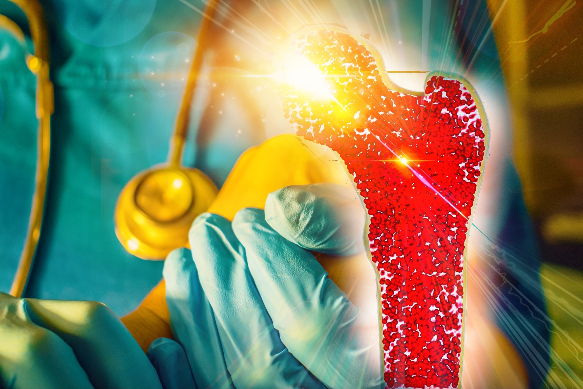
Image Credit: CI Photos / Shutterstock
Introduction
Bent bone dysplasia (BBD) is an exceptionally rare, often perinatal-lethal skeletal disorder characterized by bent long bones, osteopenia, poor calvarial mineralization, and craniosynostosis (often severe turribrachycephaly rather than isolated coronal fusion).
It most commonly results from heterozygous variants in fibroblast growth factor receptor 2 (FGFR2) that disrupt osteogenesis. Prenatally, ultrasound may reveal limb bowing, a small thorax, and craniofacial anomalies; definitive diagnosis relies on molecular testing.
Clinically, BBD is relevant for prenatal counseling, delivery planning, and neonatal care, and must be distinguished from other skeletal dysplasias to guide discussions of prognosis and recurrence risk.1
This article explains BBD, an extremely rare skeletal disorder, outlining its definition, the FGFR2 gene mutations that cause it, hallmark bent long bones, diagnosis, prognosis, and family-centered supportive care.
Definition and Key Features
BBD is a perinatal-lethal skeletal dysplasia marked by abnormal bone development with prominently bowed or bent long bones. Findings are often evident prenatally.
Core features include osteopenia; diminished calvarial mineralization with craniosynostosis; hypoplastic clavicles and pubis; narrowed ischia; bony nodules and periosteal reaction in the phalanges; and a thickened, hypercellular periosteum with smaller hypertrophic chondrocytes at the growth plate.
Distinctive radiologic hallmarks also include short, thick clavicles, wavy ribs, and striking anteroposterior vertebral shortening visible in early gestation, sometimes accompanied by femoral fractures.1,5 Craniofacial findings include hypertelorism, midface hypoplasia, micrognathia, and occasionally prenatal teeth, with a bell-shaped thorax reported.2
At the molecular level, missense substitutions in the hydrophobic transmembrane helix of FGFR2 reduce cell-surface abundance and blunt canonical fibroblast growth factor (FGF) responsiveness, establishing BBD as a distinct FGFR2-associated entity separate from other FGFR2 syndromes.
Clinically, BBD differs from collagen-defect dysplasias such as osteogenesis imperfecta and from campomelic dysplasia with SRY-box transcription factor 9 (SOX9)-related developmental anomalies. Altogether, BBD’s radiographic pattern and FGFR2 mechanism define recognizable, nosologically unique key features.2
Genetic and Molecular Basis
BBD-FGFR2 type arises from heterozygous missense mutations in FGFR2, most commonly c.1172T>G (p.Met391Arg) or c.1141T>G (p.Tyr381Asp), located within the transmembrane domain. These variants destabilize membrane insertion, reduce fully glycosylated receptor at the cell surface, sequester FGFR2 to nucleolar compartments, and markedly reduce downstream activation, including ERK1/2, consistent with deficient canonical FGF signaling. These amino acid substitutions introduce polar residues into the hydrophobic transmembrane helix, leading to mislocalization of FGFR2 within the nucleolus and diminished ligand-induced receptor phosphorylation, consistent with deficient canonical FGF signaling.2
The net result is deficient canonical FGF signaling and a disrupted balance between osteoprogenitor proliferation and differentiation, impairing both endochondral and intramembranous ossification; histology shows a hypercellular periosteum and smaller hypertrophic chondrocytes, consistent with abnormal growth-plate dynamics.1,2
Clinically, this signaling imbalance produces poor calvarial mineralization, craniosynostosis, hypoplastic clavicles/pelvis, osteopenia, and characteristically bent long bones. Inheritance is typically sporadic, with unaffected parents and de novo heterozygous FGFR2 mutations; familial genetic predisposition has not been established.
Increased intranucleolar FGFR2 activity may alter ribosomal deoxyribonucleic acid (DNA) transcription, suggesting aberrant intracellular signaling also contributes.
Rare LAMA5-related bent bone dysplasia (BBDS-2) has been described and should not be conflated with FGFR2-related BBD.1,2
Clinical Presentation
Fetal skeletal dysplasia is first suspected on routine obstetric ultrasound when long bones appear disproportionately short and show characteristic bowing or sharp angulation.
Detailed two-dimensional ultrasound (2D-US) confirms bowed or sharply angulated femora and evaluates all long bones, while three-dimensional ultrasound (3D-US) depicts whole-skeleton contours and hand/foot configuration. On ultrasound, affected fetuses may also show wavy ribs, very short clavicles, and reduced vertebral body height, features that support the early suspicion of BBD-FGFR2.1,3
Severe micromelia is common, and accompanying thoracic abnormalities, such as short ribs, a narrow chest, or platyspondyly, signal reduced intrathoracic space. These changes often correlate with pulmonary hypoplasia, the main determinant of immediate perinatal outcome, and may present clinically as respiratory insufficiency at birth.3
Ancillary findings help refine the diagnosis: frontal bossing and mid-face hypoplasia suggest FGFR3-related disorders; micrognathia points to entities such as campomelic dysplasia; a trident hand favors achondroplasia/thanatophoric dysplasia; and scapular wing hypoplasia is distinctive for campomelic dysplasia.
Spine and pelvic clues (interpedicular distance narrowing, trident ilia, delayed pubic/ischial ossification) further support classification.
Magnetic resonance imaging (MRI) can assist lung volumetry and soft-tissue assessment, and ultra-low-dose fetal computed tomography (CT) with 3D reconstruction depicts skeletal architecture when ultrasound windows are limited.
Radiographs obtained postnatally or postmortem mirror prenatal findings and may reveal fractures or metaphyseal changes that were equivocal on ultrasound, consolidating the etiologic diagnosis.3
Diagnosis
Diagnosis of BBD-FGFR2 integrates prenatal imaging and molecular confirmation, with postnatal corroboration when viable. On mid-trimester ultrasound, fetuses typically show curved/bowed long bones (often femora/tibiae), limb shortening, a narrow, bell-shaped thorax/small chest cavity, and occasionally poor calvarial mineralization; additional clues include short, thick clavicles and wavy ribs, which can be striking early, while craniosynostosis may be subtle before ossification. In early gestation, short, thick clavicles, wavy ribs, and anteroposterior vertebral body shortening are particularly suggestive and may precede overt craniosynostosis on imaging.1
Targeted genetic testing confirms pathogenic FGFR2 mutations in the transmembrane domain, most often heterozygous de novo missense variants such as c.1172T>G (p.Met391Arg) or c.1141T>G (p.Tyr381Asp), which define BBD-FGFR2.1,2
When pregnancy continues or for postnatal evaluation, radiography/CT documents bowed long bones, osteopenia, hypoplastic clavicles/pubic bones, and variable craniosynostosis; lateral spine films may reveal anteroposterior vertebral shortening.
Prenatal radiographs can corroborate the ultrasound skeletal pattern and severity. The overall clinicoradiologic pattern distinguishes BBD-FGFR2 from osteogenesis imperfecta, campomelic dysplasia, and osteoglophonic dysplasia, but definitive diagnosis rests on identifying the FGFR2 mutation.1,2
Prognosis and Outcomes
Prognosis is guarded, as many skeletal dysplasias with a markedly narrow thorax are lethal because pulmonary hypoplasia and respiratory failure occur at or shortly after birth. When identified in utero, fetal MRI-based lung volume estimation supports realistic counseling and planned delivery at a tertiary neonatal intensive care unit (NICU) prepared for high-frequency ventilation (HFV) and inhaled nitric oxide (iNO). Such preparation cannot increase intrinsic lung size but can improve immediate stabilization and, in select airway-obstruction scenarios, allow ex utero intrapartum treatment (EXIT) when upper-airway obstruction is anticipated.4
Among survivors, outcomes mirror the degree of chest wall restriction and airway disease: some require prolonged supplemental oxygen, continuous positive airway pressure (CPAP), or a heated, humidified, high-flow nasal cannula (HHFNC) for weeks to months. Because restrictive thoracic mechanics and pulmonary hypoplasia dominate morbidity, functional residual capacity is markedly reduced, and survivors may develop chronic lung disease or pulmonary hypertension.4
Ongoing risks include growth failure, sleep-disordered breathing, recurrent infections, and pulmonary hypertension, requiring structured follow-up. Multidisciplinary respiratory, cardiology, and nutrition care can enhance quality of life and extend survival.4
Management and Supportive Care
No curative treatment exists for BBD-FGFR2, as thoracic restriction and respiratory failure usually lead to perinatal lethality. Management centers on anticipatory, compassionate, supportive care. During pregnancy, team counseling should review outcomes, options for perinatal palliation, and delivery planning at a tertiary center with neonatology, genetics, and radiology.
After birth, care focuses on airway support aligned with family preferences, comfort measures, and end-of-life and bereavement support when survival is not feasible. Postnatal respiratory stabilization may require non-invasive ventilation or mechanical support for prolonged periods; management should address airway malacia and restrictive lung disease characteristic of skeletal dysplasia infants.4 Family counseling is essential to prepare parents for craniofacial differences, bowed long bones, and possible fractures, and to coordinate psychological and social support.
Genetic counseling should explain that pathogenic variants in FGFR2 are typically de novo, discuss a small yet non-zero recurrence risk from gonadal mosaicism, and outline options for future pregnancies, including targeted FGFR2 testing of chorionic villi or amniotic fluid and fetal imaging.1
Research and Future Directions
Work on BBD-FGFR2 clarifies how mutations in FGFR2 disrupt canonical FGF signaling: mutant receptors show reduced plasma-membrane trafficking, failure to activate ERK1/2, and relative nuclear retention, illuminating an imbalance in osteoprogenitor proliferation and differentiation.
These insights generalize to skeletal biology: FGFR2 is required for endochondral and intramembranous ossification; splice-variant studies (IIIb vs IIIc) map epithelial–mesenchymal roles; and pathway links span craniosynostosis syndromes and even genital tubercle development, broadening relevance beyond BBD.1,2
The largest reported cohort of 11 individuals delineated the moustache-shaped clavicles and angel-shaped phalanges as reproducible diagnostic signs and confirmed that all had heterozygous FGFR2 transmembrane mutations.5
Future work should dissect the intracellular (non-membrane) signaling pathways of mutant FGFR2 in cell and mouse models to resolve phenotypic complexity. Clinically, more detailed case series and disease registries are needed: BBD-FGFR2 may be under-recognized or misclassified, and systematic capture of radiographic hallmarks (e.g., very short clavicles, wavy ribs, vertebral shortening; occasional fractures) will refine diagnosis, natural history, and counseling.1,2
Conclusions
Early, accurate diagnosis of BBD, ideally combining targeted prenatal imaging with molecular confirmation, enables compassionate planning. Care should prioritize comfort-focused, family-centered support: coordinated delivery at centers; individualized respiratory management; pain control; and genetic counseling that explains sporadic FGFR2 variants and recurrence options for future pregnancies.
Transparent discussions about prognosis, palliative pathways, and bereavement resources uphold dignity and significantly reduce decisional burden. Although no curative therapy exists, sustained progress in genetics and developmental skeletal biology, especially deeper mapping of FGFR2 trafficking and signaling, accurate case registries, and experimental models, offers hope for sharper diagnoses and, eventually, targeted interventions.
UK's Tiniest Teen On Living With Skeletal Dysplasia | This Morning
References
- Handa, A., Okajima, Y., Izumi, N., Yamanaka, M., & Kurihara, Y. (2016). Bent bone dysplasia (BBD)-FGFR2 type: the radiologic manifestations in early gestation. Pediatric radiology. 46(2). 296-299. DOI: 10.1007/s00247-015-3465-y, https://link.springer.com/article/10.1007/s00247-015-3465-y
- Merrill, A. E., Sarukhanov, A., Krejci, P., Idoni, B., Camacho, N., Estrada, K. D., Lyons, K. M., Deixler, H., Robinson, H., Chitayat, D., Curry, C. J., Lachman, R. S., Wilcox, W. R., & Krakow, D. (2012). Bent bone dysplasia-FGFR2 type, a distinct skeletal disorder, has deficient canonical FGF signaling. The American Journal of Human Genetics. 90(3). 550-557. DOI: 10.1016/j.ajhg.2012.02.005, https://www.cell.com/ajhg/fulltext/S0002-9297(12)00090-0
- Nishimura, G., Handa, A., Miyazaki, O., Tsujioka, Y., Murotsuki, J., Sawai, H., Yamada, T., Kozuma, Y., Takahashi, Y., Ozawa, K., Pooh, R., & Sase, M. Prenatal diagnosis of bone dysplasias. British Journal of Radiology. 96 (1147). DOI: 10.1259/bjr.20221025, https://academic.oup.com/bjr/article/96/1147/20221025/7469168
- Alapati, D., & Shaffer, T. H. (2017). Skeletal dysplasia: Respiratory management during infancy. Respiratory medicine. 131. 18-26. DOI: 10.1016/j.rmed.2017.07.063, https://www.resmedjournal.com/article/S0954-6111(17)30262-7/fulltext
- Krakow, D., Cohn, D. H., Wilcox, W. R., Noh, G. J., Raffel, L. J., Sarukhanov, A., Ivanova, M. H., Danielpour, M., Grange, D. K., Elliott, A. M., Bernstein, J. A., Rimoin, D. L., Merrill, A. E., & Lachman, R. S. (2016). Clinical and radiographic delineation of Bent Bone Dysplasia-FGFR2 type or Bent Bone Dysplasia with Distinctive Clavicles and Angel-shaped Phalanges. American Journal of Medical Genetics Part A, 170(10), 2652-2661. DOI: 10.1002/ajmg.a.37772, https://onlinelibrary.wiley.com/doi/10.1002/ajmg.a.37772
Further Reading
Last Updated: Oct 23, 2025