Apr 5 2017
Large-area chemical imaging reveals original paint layers on Ghent altarpiece
Considered the pinnacle of mediaeval painting, the Ghent Altarpiece was painted around 1432 by Jan van Eyck and probably his brother Hubert. It is currently undergoing the most extensive conservation treatment for more than a century. The decision to remove all overpaint was underpinned by scientific arguments: Belgian researchers report in the journal Angewandte Chemie about their use of “chemical maps” to visualize the original paint layers under the overpainted surface.
Having survived an eventful history, the 4-meter-wide and over 3-meter-high winged altar stands in the Cathedral of St. Bavo in Ghent, Belgium, where it is viewed by about 200,000 visitors every year. This work of art has already been restored several times, but a new, stepwise restoration was begun in 2012. Belgian scientists at the University of Antwerp, the Royal Institute for Cultural Heritage (KIK-IRPA, Brussels), and the University of Ghent, analyzed the scenes on the backside of the eight wing panels , which are visible when the altar is closed. They used chemical imaging techniques based on element-specific X-ray analysis to adjust and optimize the conservation strategy and to monitor the removal of the overpaint during the cleaning phase.
Previously, it was only possible to examine works of art point by point, which is not very representative. Led by Geert Van der Snickt, the team used mobile scanning systems to image the entire surface by X-ray fluorescence spectroscopy. In this method, X-rays knock electrons out of the inner shells of the atom. When electrons from outer shells fill these empty spots, they release energy as element-specific X-ray fluorescence.
“More than 16 million spectra were collected and swiftly processed with our in-house software to yield over 1 GB of spectral data for each panel,” says Van der Snickt. “By using computer calculations, we were able to translate these into chemical maps that depict the distribution of elements. X-rays penetrate the layers of paint without damaging them. In this way, we were able to visualize the original Van Eyck paint layers hidden under the overpaint.” To gain more information about the detailed structure of the layers, the researchers also analyzed cross-sections of tiny samples of paint.
Says Van der Snickt:
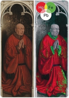
For example, in an analysis of the portrait of kneeling donor Joos Vyd, the lead, mercury, and iron maps revealed substantial damage to the original paint in an area of the vermillion red robe that appeared undamaged. It also showed how the gaps were filled with a red iron-containing paste before being covered with a thin layer of red mercury sulfide paint.
However, by revealing the overall good condition of the original scenes, the chemical maps supported the decision to remove all overpaints that were previously thought to be Van Eyck’s work.
 AI maps how brain fluids move to clear waste