A mouse study maps two specialized nerve cell populations within the heart, revealing how one supports everyday cardiac function while the other helps prevent electrical collapse during severe stress.
Study: The intrinsic cardiac nervous system is essential for cardiac function and survival
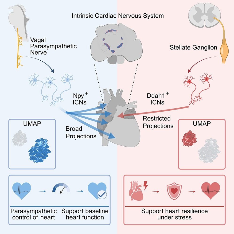
A recent mouse study in the journal Cell suggests that the intrinsic cardiac nervous system (ICNS) is vital for proper functioning of the mouse heart. Integrating genetic, imaging, and cell-specific neuromodulation studies in adult mice, researchers identified two distinct types of intrinsic cardiac neurons (ICNs), one crucial for regulating heart rate and supporting coronary perfusion and the other for maintaining electrical stability during extreme stress. Further mechanistic investigations into the ICNS structure and function could inform future cell-type-targeted neuromodulatory therapies, although the human relevance of these findings remains unknown.
The ICNS is a key node in communication between the heart and the brain and is implicated in several cardiac disorders. However, its functional organization remains poorly understood because most studies have primarily focused on extrinsic autonomic circuits. Currently used therapies, including radiofrequency ablation, electrical stimulation, and cryoablation, broadly target elements of the cardiac nervous system without cell-type specificity and often produce variable outcomes. An improved understanding of the ICNS could help researchers design more targeted treatments in the future, although clinical benefit in humans remains untested.
About the Study
In the present study, researchers integrated genetic investigations with imaging methods in adult mice to investigate the role of the ICNS in cardiac function. They performed single-cell RNA sequencing (scRNA-seq), genetic labeling studies, high-resolution volumetric imaging, and cell-specific neuromodulation studies to dissect the complex ICNS framework. To characterize ICN subtypes, the team performed adeno-associated virus (AAV)-based genetic labeling and mapped neuronal morphology and cell-type-specific innervation patterns.
The researchers developed genetically engineered mouse models to selectively label, target, and manipulate specific cell types in the ICNS. They performed fluorescence-activated cell sorting (FACS) to purify green fluorescent protein-labeled (GFP) ICNs from anterior, posterior, and mixed cardiac regions. RNAscope analysis validated the single-cell findings and confirmed the distributions in situ.
The team identified molecularly distinct ICN subtypes. These populations received different inputs from extrinsic circuits and innervated distinct domains of the cardiac system to perform different functions. They also performed diphtheria toxin (DT)-mediated ablation experiments and assessed the effects. Single-cell RNA sequencing of 2,870 ICNs identified four transcriptional clusters, visualized using uniform manifold approximation and projection (UMAP), with Npy-positive and Ddah1-positive cells constituting the two major molecular classes. They also subjected the animals to physical restraint, sustained heat exposure, and epinephrine-caffeine-induced sympathetic overactivation to study ICNS function under stressful conditions.
Results
The ICNS lies at the interface between extrinsic autonomic inputs and cardiac cells, positioning it to integrate local and central signals. Through genetic labeling, the researchers identified GFP-positive ICNs situated across several anterior and posterior ganglia on the atrium. Most ICNs were cholinergic. The team found that neurons expressing neuropeptide Y (Npy) and dimethylarginine dimethylaminohydrolase 1 (Ddah1) were the two major molecular subtypes in the ICNS. These ICNs differed in neural inputs and cardiac projections. Npy-positive ICNs preferentially received vagal input, regulated heart rate through the parasympathetic nervous system, and supported coronary perfusion by modulating aortic-root mechanics. Ablation of Npy-positive ICNs led to fatal cardiac failure in mice.
Ddah1-positive ICNs, enriched in the posterior ganglia, received sympathetic inputs and were found to be crucial for maintaining cardiac electrical stability under extreme physiological or psychological stress. Their ablation increased susceptibility to malignant arrhythmias and sudden cardiac arrest. Activation of these ICNs exerted cardioprotective functions by improving survival during epinephrine-caffeine-induced sympathetic overactivation in mice.
Ddah1-positive neurons frequently co-expressed dopamine beta-hydroxylase (Dbh), but this molecular feature did not establish a classical sympathetic identity. Separate monosynaptic tracing showed that Ddah1-positive ICNs received direct sympathetic input. Npy-positive neurons frequently expressed prostaglandin E receptor 3 (PTGER3), indicating a distinct molecular subtype whose functional relevance was not tested. Npy-positive ICNs most closely resembled inhibitory motor neurons in the gut at the transcriptomic level, although their precise downstream effects were not tested. Ddah1-positive ICNs shared transcriptomic features with certain sensory- and interneuron-like populations in the gut, but their sensory modalities and circuit functions remain unknown.
Npy-positive ICN projections extensively innervated the heart, reaching the atrial appendages, ventricular tissue, cardiac apex, and pulmonary venous region. Npy-positive fibers targeted the atrioventricular (AV) and sinoatrial (SA) nodes, suggesting that these ICNs modulate cardiac rhythm. They also densely encircled the root of the aorta, suggesting roles in regulating cardiac outflow. In contrast, Ddah1-positive ICNs showed restricted innervation confined to the left atrial tissues, pulmonary veins, and pulmonary artery.
In particular, these ICNs targeted the openings where the pulmonary veins enter the left atrium, a region associated with atrial fibrillation and arrhythmias. Ddah1-expressing ICNs also displayed relatively sparse local projections but exhibited a more extensive network of nerve fibers linking ICN ganglia. The findings are consistent with spatially limited communication between these ICNs and cardiac targets but also suggest a potential role in local circuit integration.
Conclusions
The findings demonstrate that the ICNS neural network is critical for regulating cardiac function in mice and could ultimately help guide more precise, cell-type-targeted neuromodulation, although therapeutic benefit in humans has not been established. Future studies in mice should use single-neuron tracing and cell-specific functional mapping to determine whether individual Npy-positive neurons are multifunctional or whether the Npy-positive population comprises specialized subgroups. Future studies should also clarify the precise mechanism of action, sensory modalities, and downstream targets of Ddah1-positive ICNs and determine whether the molecular and functional organization of these ICNs is conserved in the human heart.
 Drug-coated balloons offer an alternative to permanent heart stents