MR Solutions, with offices in the UK and Boston, will be showing their latest state of the art 3T to 9.4T superconducting, cryogen-free MRI imaging technology with optional PET and SPECT imaging modalities on stand 407 at the 16th annual World Preclinical Congress in Boston on June 13th to 15th. The congress focusses on the very latest trends and technologies impacting the preclinical drug discovery and development world.
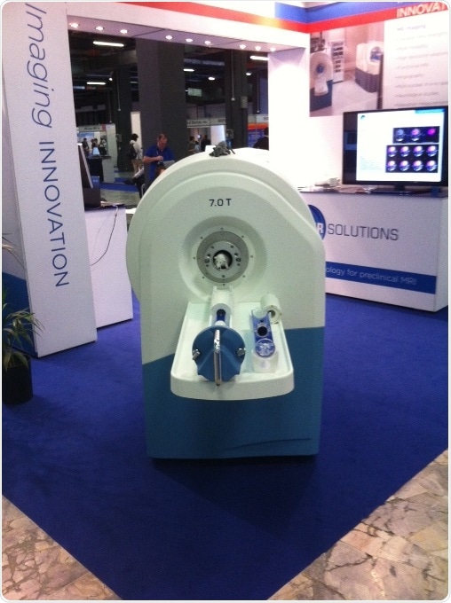
7T Cryogen Free preclinical MRI System - MRS 7000 Series
MR Solutions’ preclinical imaging systems are revolutionising drug discovery by providing better scanners with a more viable footprint by doing away with all the equipment and building modifications required for liquid helium cooled machines. The company is at the forefront of preclinical MRI, transforming scanners which previously required a whole room into a powerful, benchtop unit which can simply be wheeled in to the laboratory.
Physicist and CEO of MR Solutions Dr David Taylor said:
Our preclinical MRI imaging systems which will be on display at the World Preclinical Congress are now changing the way that new candidate drugs are being developed. These systems are the link between visualisation at molecular level and translational drug development.
MR Solutions was able to develop an MRI system using a novel superconducting magnet design which only needs a readily available cryocooler unit to get to the very low temperature required. MR Solutions’ 3T to 9.4T MRI benchtop scanners are used in research laboratories across the world and can be installed alongside other equipment because the innovative design has reduced the stray magnetic field from metres to centimetres meaning that it does not interfere with other laboratory equipment.
For simultaneous imaging, a PET or SPECT imaging module can be inserted within the magnet of MR Solutions’ MRI system. For sequential imaging, a PET or SPECT imaging module can be clipped on to the front of the MRI system. This allows imaging to be carried out automatically from one modality to another on the same axis.
 Aircraft emissions could double air pollution deaths by 2040