The spike (S) protein of the severe acute respiratory syndrome coronavirus 2 (SARS-CoV-2) plays a critical role in receptor recognition and host-cell entry. Thus, major efforts to characterize and reproduce this protein in its truest form have been underway since the inception of the coronavirus disease 2019 (COVID-19) pandemic to support the development of adequate diagnostic and therapeutic measures.
The receptor-binding domain (RBD) of the S protein is responsible for interaction with the angiotensin-converting enzyme 2 (ACE2) receptor of the host cell. Therefore, accurate reproduction of this region is vital to the outcome of serological tests, the development of vaccines, and drug discovery efforts.
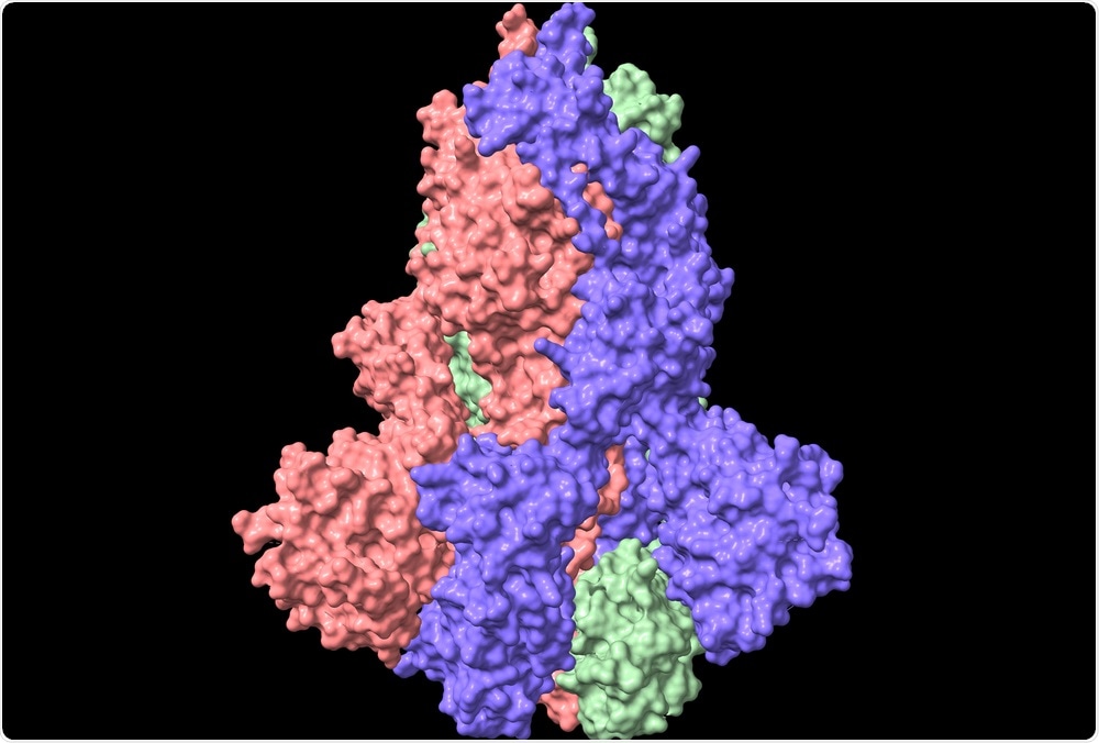 Study: The nuts and bolts of SARS-CoV-2 Spike Receptor Binding Domain heterologous expression. Image Credit: Volodymyr Dvorynk / Shutterstock.com
 This news article was a review of a preliminary scientific report that had not undergone peer-review at the time of publication. Since its initial publication, the scientific report has now been peer reviewed and accepted for publication in a Scientific Journal. Links to the preliminary and peer-reviewed reports are available in the Sources section at the bottom of this article. View Sources
Mammalian, insect, and bacterial cells have been utilized to heterologously produce recombinant SARS-CoV-2 RBD proteins by a variety of methods. In a recent study published on the bioRxiv* preprint server, RBD produced in each of these cell types are characterized in-depth, comparing their applicability for future COVID-19 research.
RBD generation
In the current study, the SARS-CoV-2 RBD flanked by a His-tag was cloned in mammalian HEK-293 cells. Cells were then transfected with deoxyribonucleic acid (DNA) and left to produce the protein for one week.
The medium containing the RBD was then filtered and purified by affinity chromatography, and the eluted protein was confirmed by anti-His and anti-S protein antibodies. The molecular mass of the RBD was higher than expected, thereby suggesting that post-translational modifications of the protein had taken place within the cells.
RBD generation in insect cells was achieved firstly by generation and amplification of baculovirus in Sf21, and subsequent RBD protein expression in infected Hi-5 cells. Proteins of interest were separated from the cell culture media and analyzed by affinity chromatography. In this experiment, the researchers found that the proteins bore a closer molecular weight to the true RBD as compared to those produced in mammalian cells, while also achieving a higher purity of 95%.
The RBD produced by two different strains of Escherichia coli was also characterized. The first strain included BL-21 Star, which is commonly employed to generate high levels of expression of non-toxic recombinant proteins. The second strain was Lemo21, which allows for the expression of challenging toxic or insoluble proteins by limiting inclusion body formation.
The RBD protein was then collected from the inclusion bodies of E. coli at yields of 5.2% and 8.1% for BL-21 Star and Lemo21 strains, respectively. A purity of 90% was achieved by dialysis in the presence of denaturing agents. The final proteins demonstrated a good correlation with the true molecular weight of the SARS-CoV-2 RBD.
Comparing RBDs
While the RBD proteins produced by E. coli eluted as a single narrow peak during size exclusion chromatography, those generated in mammalian and insect cells demonstrated shifts in their elution peak that suggest the presence of extensive glycosylation. E. coli produced RBD eluted at 18.7 mL, whereas mammalian cells produced RBD with an elution peak at 15.3 mL. Comparably, insect RBDs eluted at both 16 and 14.6 mL, thus indicating that two distinct populations with differing glycosylation patterns were present in these cells.
The conformational stability of each RBD was also tested using temperature destabilization experiments. Here, the researchers found that each protein was stable to around 323 K with some minor variation, matching reports of the thermal stability of the native SARS-CoV-2 RBD.
Interestingly, the researchers of the current study note biphasic behavior in the unfolding of the E. coli produced RBD. More specifically, an initial small unfolding event is detected at 305 K before the main body of the protein is denatured at 307 K. The group speculates that this is likely due to a failure in protein unfolding during production, owing to the lack of glycosylation on these proteins.
The affinity of commercially available RBD antibodies and rat immunoglobulin G (IgG) generated by vaccination towards each RBD was assessed by enzyme linked immunosorbent assay (ELISA). Through the use of this assay, the researchers found that both insect and mammalian-produced RBDs were efficiently recognized, while E. coli produced RBDs exhibited a much lower affinity.
Against Vero-E6 cells expressing the ACE2 receptor, all three RBDs demonstrated similar efficacy in recognizing the receptor. However, the affinity between ACE2 and the E. coli-generated RBD was shown to be significantly lower than towards those generated in insect or mammalian cells using bio-layer interferometry, with the latter achieving the greatest affinity.
(a) Serum from immunized rat with COVID-eVax was used to compare different concentrations (1, 3, 5 μg/mL) of RBD expressed in E. coli(green), insect (blue) and HEK-293 cells (black). The y-axis represents the optical density (OD) measured at 405 nm while the x-axis accounts for RBD concentrations and serum dilution factors (1:1000, 1:10000, 1:50000). Bars indicate standard deviations. (b) Commercial antibody against the S1 subunit of SARS-CoV-2 Spike was used to compare different concentrations (1, 3, 5 μg/mL) of RBD produced in E. coli (green), insect (blue) and HEK-293 cells (black). Optical density (OD) was measured at 450 nm and bars indicate standard deviations.
The yields of RBD generated were up to 60 mg/liter of culture for mammalian and insect cells, though they were highly glycosylated. In contrast, E. coli yields were significantly lower and the generated proteins lacked glycosylation, which is a major factor in the interaction of the protein with antibodies and the ACE2 receptor that likely contributed towards the observed lower affinity.
However, the production time of E. coli RBD is less than 1 week. Comparably, generation by insect or mammalian cells requires 2-3 weeks and entails significantly higher production cost. Thus, E. coli RBD may be suitable for some specific research purposes where accurate glycosylation is essential.
This news article was a review of a preliminary scientific report that had not undergone peer-review at the time of publication. Since its initial publication, the scientific report has now been peer reviewed and accepted for publication in a Scientific Journal. Links to the preliminary and peer-reviewed reports are available in the Sources section at the bottom of this article. View Sources
Journal references:
- Preliminary scientific report.
Maffei, G., Montemiglio, L. C., Vitagliano, G., et al. (2021). The nuts and bolts of SARS-CoV-2 Spike Receptor Binding Domain heterologous expression. bioRxiv. doi:10.1101/2021.09.17.460782. https://www.biorxiv.org/content/10.1101/2021.09.17.460782v1
- Peer reviewed and published scientific report.
Maffei, Mariano, Linda Celeste Montemiglio, Grazia Vitagliano, Luigi Fedele, Shaila Sellathurai, Federica Bucci, Mirco Compagnone, et al. 2021. “The Nuts and Bolts of SARS-CoV-2 Spike Receptor-Binding Domain Heterologous Expression.” Biomolecules 11 (12): 1812. https://doi.org/10.3390/biom11121812. https://www.mdpi.com/2218-273X/11/12/1812.
 Scientists develop a dual action strategy to destabilize the COVID virus