New research reveals how fine-tuning retinoic acid metabolism enables the axolotl to regrow complex limbs, opening up new possibilities for regenerative medicine.
 Study: Retinoic acid breakdown is required for proximodistal positional identity during axolotl limb regeneration. Image Credit: Bigzumi / Shutterstock
In a recent study published in the journal Nature Communications, researchers in the United States explored how cytochrome P450 family 26 subfamily B member 1 (CYP26B1)-mediated retinoic acid (RA) breakdown regulates proximodistal (PD) identity during axolotl limb regeneration.
The study also addresses unresolved questions regarding how RA gradients are established during regeneration and how these mechanisms might translate into future applications for regenerative medicine.
Background
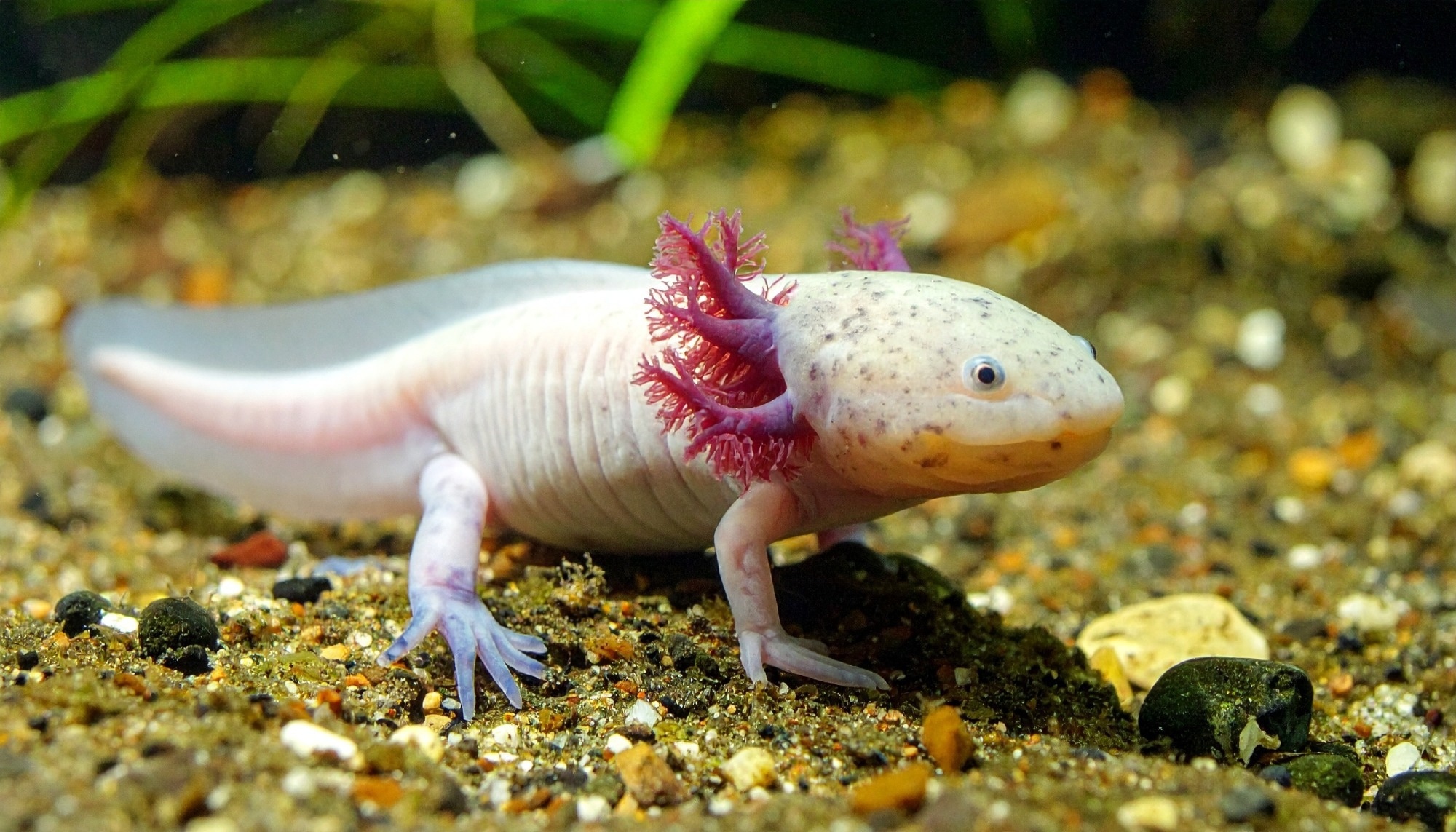
Imagine losing a limb and watching it grow back perfectly, bone by bone and joint by joint. While this sounds like science fiction for humans, axolotls (a type of salamander) do it routinely. They regenerate limbs with remarkable precision, restoring the correct segments from the shoulder (stylopod) to the hand (autopod). This ability hinges on the blastema, a cluster of stem-like cells that “remember” their original position. RA influences this memory, but how its levels are controlled during regeneration remains unclear. Understanding these controls could transform regenerative medicine. Further research is needed to decode the molecular mechanisms guiding positional identity. The authors also note that while the importance of RA in positional identity is established, whether RA synthesis is graded along the limb axis or is uniform and instead controlled by localized degradation remains an open question.
About the study
The study utilized axolotls with limbs amputated at defined proximal-distal (PD) levels, including upper stylopod (US), lower stylopod (LS), upper zeugopod (UZ), and autopod (A), to investigate RA signaling. Quantitative reverse transcription polymerase chain reaction (qRT-PCR), hybridization chain reaction fluorescence in situ hybridization (HCR-FISH), and single-cell Ribonucleic Acid sequencing (scRNA-seq) were used to assess gene expression. The enzymes and genes involved in RA synthesis, including retinaldehyde dehydrogenase 1 (RALDH1), RALDH2, and RALDH3, RA degradation, such as CYP26A1 and CYP26B1, and RA response, including retinoic acid receptor alpha (RARA) and retinoic acid receptor gamma (RARG), were profiled across proximal blastemas (PBs) and distal blastemas (DBs).
To manipulate RA levels, animals were treated with talarozole (TAL, also known as R115866), an inhibitor of CYP26 enzymes, at concentrations of 0.1 micromolar (μM), 1 μM, and 5 μM. RA activity was measured using RA-responsive element: enhanced green fluorescent protein (RARE:EGFP) reporter animals. In some experiments, TAL was combined with disulfiram (DIS), a pan-RALDH inhibitor, or AGN 193109 (RAA), a pan-retinoic acid receptor (RAR) antagonist. The expression of patterning genes Meis homeobox 1 and 2 (Meis1, Meis2), homeobox A cluster genes (Hoxa9, Hoxa11, Hoxa13), and short stature homeobox (Shox, Shox2) was evaluated. Clustered Regularly Interspaced Short Palindromic Repeats / CRISPR-associated protein 9 (CRISPR/Cas9) was used to generate Shox knockout animals (Shox−/−) to assess its functional role.
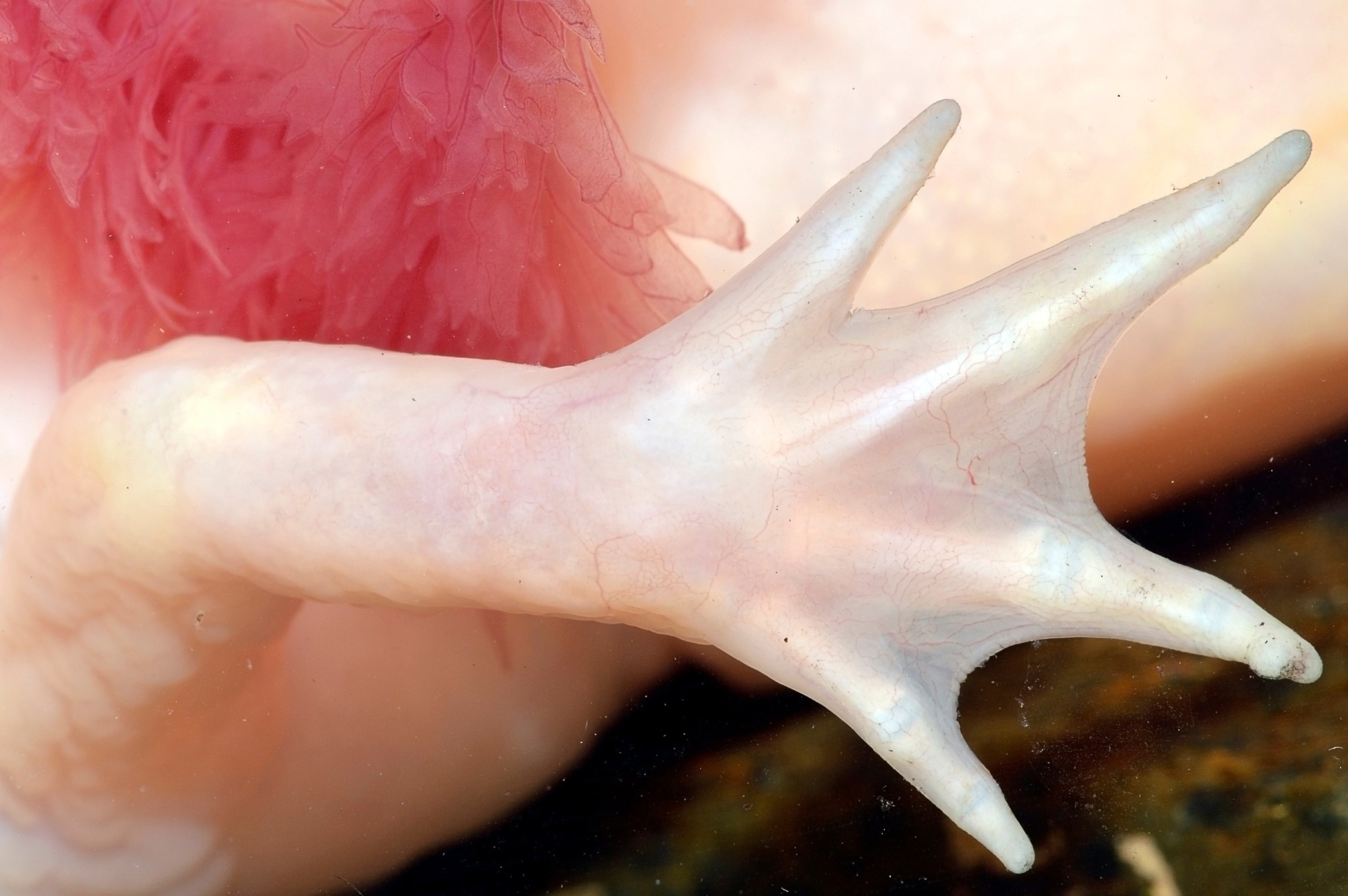
 The forelimb of Ambystoma mexicanum (axolotl, albino variant), a neotenic salamander (Ambystomatidae). Image Credit: Guillermo Guerao Serra / Shutterstock
Study results
The study confirmed that RA signaling varies along the PD axis, with higher levels in PBs compared to DBs. CYP26B1 expression was significantly higher in the mesenchyme of DBs. In contrast, Meis1 and Meis2, which are RA-responsive genes linked to proximal identity, were enriched in PBs. Hoxa13 was highly expressed in DBs. These expression patterns matched limb segment positions and were evolutionarily conserved.
Inhibiting RA degradation with TAL increased RA signaling, as visualized by RARE:EGFP reporters, and induced proximal limb duplications from distal amputations. TAL treatment caused DBs to regenerate stylopods or zeugopods instead of autopods. At 1 μM TAL, 66.7% of DBs developed full stylopod duplications. In contrast, proximally amputated limbs treated with the same TAL concentration regenerated normally, although higher doses impaired regeneration. These effects confirmed that CYP26B1 regulates RA concentrations critical for segment-specific patterning.
TAL-treated DBs showed reduced Hoxa13 and increased Meis1 expression. RNA sequencing revealed a dose-dependent transcriptional reprogramming, with TAL-treated DBs adopting expression profiles similar to dimethyl sulfoxide (DMSO)-treated PBs. Among differentially expressed genes were Shox and Shox2, which are RA-responsive and enriched in PBs. Shox expression was inversely correlated with Hoxa13 and co-localized with Meis1, suggesting it acts downstream of RA signaling and Meis1. The authors note that Meis1 is likely upstream of Shox in this regulatory cascade, supported by promoter analysis, and that Shox and Hoxa13 expressing cells were mutually exclusive.
Functional studies using CRISPR/Cas9-induced Shox knockout animals revealed that Shox−/− axolotls developed shortened stylopods and zeugopods, with impaired endochondral ossification. Histological staining and hematoxylin and eosin analysis showed immature chondrocytes that failed to mature into bone. However, the autopod remained unaffected. Despite these skeletal defects, Shox−/− animals regenerated limbs, confirming that Shox is not essential for regeneration but is crucial for patterning proximal skeletal segments. The study also observed that Shox2 expression can partially compensate for loss of Shox in some contexts, although proximal defects persist, and that chondrocyte formation in distal elements is Shox-independent.
Shox expression was predominantly observed in mesenchymal cells and was activated in TAL-treated DBs. When TAL was co-administered with either DIS or RAA, the proximalization of DBs was blocked, showing that both RA synthesis and receptor-mediated signaling are required for this effect. Furthermore, in Shox−/− animals treated with TAL, limb duplications occurred, but the duplicated segments were underdeveloped, indicating that Shox is necessary to execute, but not initiate, RA-induced proximal patterning.
While the study presents strong evidence that Cyp26b1-mediated RA degradation controls segmental identity, it also discusses that the upstream regulators of Cyp26b1 (such as Hoxa11, Hoxa13, or FGF signals) during regeneration remain to be determined.
Conclusions
To summarize, this study demonstrates that CYP26B1-dependent retinoic acid (RA) degradation is crucial for the establishment of PD identity during axolotl limb regeneration. By adjusting RA levels in the blastema, CYP26B1 enables segment-specific activation of patterning genes. Elevated RA levels promote proximal identity through Meis1 and Shox, while RA degradation supports distal identity marked by Hoxa13. Shox is critical for the formation and ossification of stylopodial and zeugopodial elements but is not required for autopod formation or overall regeneration.
The findings are also consistent with a segment-specific threshold model for positional identity (similar to the French Flag model), though the paper highlights uncertainties regarding whether RA synthesis itself is uniform or graded along the PD axis. These insights underscore the importance of localized RA metabolism in regenerating complex structures and may inform future regenerative strategies in medicine and developmental biology.
Journal reference:
- Duerr, T.J., Miller, M., Kumar, S. et al. Retinoic acid breakdown is required for proximodistal positional identity during axolotl limb regeneration. Nat Commun (2025), DOI: 10.1038/s41467-025-59497-5, https://www.nature.com/articles/s41467-025-59497-5